Hep B Blog

Category Archives: Hepatitis B Research

Hepatitis B Research Review: May

This month, research from Melbourne, Australia indicates that the kinases TBK1 and IKKε act redundantly to initiate STING-induced, NF-kB-mediated transcription of proinflammatory cytokines. Nearby researchers also working in Melbourne have demonstrated that an HBV vaccine composed of glycosylated HBV surface protein outperforms those currently in use.  Also, researchers at St. Jude Children’s Research Hospital in Memphis, Tennessee have elucidated the role of caspase-6 in influenza A virus host defense.
  • TBK1 and IKKε Act Redundantly to Mediate STING Induced NF-kB Responses in Myeloid Cells – Cell Reports
    • This paper from The Walter and Eliza Hall Institute of Medical Research in Melbourne, Australia deciphers the role of the kinases TBK1 and IKKε in STING-induced, NF-kB-mediated cytokine production. Stimulator of Interferon Genes (STING) protein is a vital component of the innate immune system. Cyclic guanosine monophosphate-adenosine monophosphate (cGAMP) synthase (cGAS), is a pattern recognition receptor (PRR) that senses cytoplasmic double-stranded DNA (dsDNA). In response to dsDNA binding, cGAS catalyzes the production of 2’3′-cGAMP, a cyclic dinucleotide (CDN) which activates STING by direct binding. Once bound to 2’3′-cGAMP, STING dimers undergo a conformational change and translocate from the endoplasmic reticulum (ER) to the Golgi apparatus. At the Golgi, the serine-threonine protein kinase TANK-binding kinase 1 (TBK1) phosphorylates STING at residues in its C-terminal tail (CTT). This phosphorylation causes the recruitment of interferon regulatory factor 3 (IRF3) to STING which is also phosphorylated by TBK1. Phosphorylated IRF3 forms dimers and translocates to the nucleus where it induces the expression of type I interferons (IFN-I) such as IFN-β. IFN-I production and secretion lead to the activation of numerous IFN-stimulated genes (ISGs) which induce a robust antiviral state in the cell. Concomitant to IFN-I induction, STING activation is also known to induce a set of proinflammatory cytokines through the transcription factor called nuclear factor-kB (NF-kB). These cytokines include tumor necrosis factor alpha (TNFα) and interleukins (IL) IL-1β and IL-6. While TBK1 and to a much lesser extent IkB kinase ε (IKKε) are needed for IRF3-mediated IFN-I transcription, several lines of evidence indicate that they may be unnecessary for STING-induced NF-kB activity. For instance, the CTT region of STING, critical to IFN induction, is observed only in vertebrates. While STING activation in the invertebrate species Drosophila melanogaster and Nematostella vectensis results in NF-kB-mediated transcription of cytokines, it does not induce IFN-I transcription. Additionally, ubiquitination of STING at lysine residues K244 and K288 which is required for its trafficking from the ER to the Golgi is essential for IFN-I induction, but not for NF-kB activation. Finally, phosphorylation of STING at serine residues S358 and S366 in the CTT is required for IRF3 activation but is unnecessary for NF-kB activity. This publication reports that while TBK1 kinase activity is critical for IRF3 activation, TBK1 and IKKε act redundantly and in a kinase-independent manner to activate NF-kB signaling. To determine this, conditional TBK1-knockout mice were generated. These mice were the offspring of mice “floxed” for TBK1 and “RosaCre” mice (ROSA26-CreERT2). The floxed mice were mutated to have their TBK1 gene sandwiched between two lox P sites (Tbk1fl/fl). The RosaCre mice were mutated to constituatively produce a fusion protein of the Cre recombinase and the estrogen receptor (CreER).  The TBK1 conditional knockout mice (Tbk1fl/fl x RosaCre) transcribe TBK1 until they are treated with the synthetic steroid tamoxifen. Tamoxifen binds the the CreER fusion protein (CreERT) and causes its translocation to the nucleus where it binds to lox P sites and its recombinase activity causes the deletion of the TBK1 gene. Conditional knockout mice had to be used to study TBK1 because complete constituative TBK1 knockout is lethal to mice. Primary bone marrow-derived macrophages (BMDM) were obtained from both tamoxifen-treated wild-type Tbk1fl/fl (WT) and Tbk1fl/fl x RosaCre (TBK1 knockout) mice. When subjected to the STING agonist 2’3′-cGAMP, BMDMs from WT mice showed phosphorylation of IRF3 by Western blot and secretion of IFN-β by ELISA. Under the same treatment, BMDMs derived from TBK1 knockout mice showed drastically reduced IRF3 phosphorylation and IFN-β secretion. Interestingly, BMDMs derived from both WT and TBK1 knockout mice secreted similar levels of TNFα when treated with 2’3′-cGAMP. Next, BMDCs from normal mice were immortalized and CRISPR/Cas9 was used to knockout expression of TBK1, IKKε, or both. Significantly, while TNFα secretion upon 2’3′-cGAMP treatment was modestly reduced by the knockout of either TBK1 or IKKε, it was almost completely ablated by the knockout of both genes. Interestingly, knockout of both genes had no effect on the secretion of TNFα in response to treatment with lipopolysaccharide (LPS). Finally, in order to determine the upstream signaling responsible for STING-mediated NF-kB activity, two proteins were investigated: transforming growth factor b-activated kinase 1 (TAK1) and inhibitor of nuclear factor kappa-B kinase subunit beta (IKKβ). Small molecule inhibitors were used to inhibit TAK1 and IKKβ prior to treatment with the mouse STING agonist DMXAA. Inhibition of both TAK1 and IKKβ resulted in diminished NF-kB activity, implicating their role as kinase activators of NF-kB downstream of STING. Taken together, these results indicate that TBK1 and IKKε act redundantly to carry out STING-mediated NF-kB activity. Additionally, it is likely that TAK1 acts downstream of TBK1 and IKKε to activate the IKK complex, resulting in NF-kB activity. This finding has direct therapeutic significance for STING-driven autoimmune disorders such as chronic polyarthritis. Many strategies for overcoming such diseases only target the IFN-I-producing pathway, while pro-inflammatory cytokine production may go unchecked. This finding elucidates a less-studied arm of STING signaling which is important for basic science and future therapies.
  •  Glycoengineered Hepatitis B Virus-Like Particles with Enhanced Immunogenicity – Vaccine
    • This paper from the Royal Melbourne Institute of Technology University in Melbourne, Australia shows that an HBV vaccine using glycosylated HBV surface protein may have better efficacy than the current vaccine. HBV encodes three surface proteins (large, medium, and small) which are truncated forms of the same protein. The small HBV surface protein (HBsAgS) contains the major antigenic determinants of the protein. In the absence of other viral proteins, HBsAgS will self-assemble into non-infectious particles termed subviral particles (SVP), also known as virus-like particles (VLP). VLPs are the major species of HBV viral particle secreted from infected hepatocytes. When grown in mammalian cells in vivo, approximately half of HBsAgS molecules receive N-glycosylation at asparagine residue N146. N-glycosylation is the addition of an oligosacharide molecule to the nitrogen atom of an asparagine residue within a protein. These modifications occur in the endoplasmic reticulum (ER) and are important for the function of proteins and for signaling within the cell. The current HBV vaccines are composed of HBsAgS VLPs grown in yeast. In contrast to VLPs grown in mammalian cells, yeast-derived VLPs have no N-glycosylation. Additionally, HBV vaccines contain adjuvants which aid in immune system stimulation. The widely-used HBV vaccines Engerix-B (GlaxoSmithKine) and Recombivax HB (Merck) contain the adjuvants aluminum hydroxide and aluminum hydroxyphosphate respectively. Aluminum salts stimulate the immune system by causing activation of the NLR family pyrin domain-containing protein 3 (NLRP3) inflammasome pathway. Upon vaccination, aluminum salt crystals are taken into local dendritic cells via phagocytosis where they rupture the lysosome, causing activation of the NLRP3 inflammasome which includes active caspase 1. The catalytic activity of caspase 1 cleaves pro-interleukin 1β (IL-1β) as well as gasdermin D into their active forms. Cleaved gasdermin D forms pores in the cell membrane resulting in the rapid release of pro-inflammatory IL-1β and ultimately causing pyroptosis, an immunogenic form of cell death. This publication shows that using glycosylated HBsAgS VLPs in the presence of aluminum hydroxide may result in a more immunogenic vaccine than that which is currently used. To study the effect of HBsAgS glycosylation, first N-terminal FLAG-tagged wild-type (WT) HBsAgS and point-mutated variants were expressed in HEK 293 cells. Variants used were threonine-to-asparagine mutant T116N and asparagine-to-glutamine mutant N146Q. The T116N mutant contained an additional asparagine available for glycosylation on the domain of HBsAgS which faces the lumen of the ER. On the other hand, the N146Q mutant lacked the asparagine which is typically N-glycosylated. SDS-PAGE followed by Coomassie staining revealed that about 50% of WT HBsAgS was glycosylated, running as two distinct bands at 27 kDa (glycosylated) 24 kDa (non-glycosylated).  However, HBsAgS mutant T116N ran as two predominant bands at 27 kDa (monoglycosylated) and 29 kDa (diglycosylated). HBsAgS mutant N146Q ran as a single band at 24 kDa, indicating no glycosylation. This result confirmed that about half of HBsAgS produced in mammalian cells are N-glycosylated at N146 and no other amino acid. Both HBsAgS mutants formed VLPs similar to WT as viewed by transmission electron microscopy. VLPs were mostly spherical with some elongated in shape. Next, following removal of N-glycans using the enzyme peptide:N-glycosidase F (PNGase), quantitative N-glycome profiling was conducted using an advanced spectrometry technique called porous graphitized carbon liquid chromatography-electrospray ionization-tandem mass spectrometry (PGC-LC-ESIMS/MS). The T116N mutant was found to have a greater N-glycan density than WT HBsAgS, but a similar distribution of N-glycan types. Finally, the immunogenicity of glycoengineered HBsAg was tested using a mouse model of vaccination. BALB/c mice were immunized at weeks 1, 3, 5, and 7 with purified WT or T116N HBsAgS in the presence or absence of aluminum hydroxide. Some mice were immunized with Engerix-B as a control group. Serum samples were taken at weeks 2, 4, 6, 8, and 18 post-vaccination and analyzed by an ELISA assay against yeast-derived VLPs. Mice immunized with T116N HBsAgS combined with aluminum hydroxide had the highest titer of anti-HBsAgS antibodies at every time point tested. This indicates that hyper-glycosylated HBsAg is more effective than non-glycosylated HBsAg in mounting an immune response. The authors propose that hyper-glycosylated HBsAgS is more readily taken into antigen-presenting cells (APCs) due to an increased affinity for manose-binding lectin receptors expressed on those cells. Additionally, hyper-glycosylation of HBsAgS may lower its strength of adsorption with aluminum hydroxide, making it more prone to release and antigen processing. Taken together, these results demonstrate that glycoengineered HBsAgS formed VLPs and when combined with aluminum hydroxide, exhibited increased immunogenicity in BALB/c mice in comparison to a currently used vaccine. This publication shows one way in which molecular cloning techniques may be used to improve the efficiency and reliability of HBV vaccines.
  • Caspase-6 Is a Key Regulator of Innate Immunity, Inflammasome Activation, and Host Defense – Cell
    • This paper from St. Jude Children’s Research Hospital in Memphis, Tennessee shows that caspase-6 mediates inflammasome activation and plays a role in the activation of the programmed cell death (PCD) pathways pyroptosis, apoptosis, and necroptosis (PANoptosis). The caspase family of proteins are cysteine-aspartic proteases which cleave proteins between cysteine and aspartic acid residues. Caspases play essential rolls in inflammation and PCD pathways. Caspases exist as inactive zymogens (pro-forms) within the cell until they are cleaved, resulting their active form. Caspases are grouped as being either inflammatory (caspase-1, -4, -5, and -11) or apoptotic (caspase-3, -6, -7, -8, -9 and -10). However, emerging evidence has demonstrated crosstalk between these groups under certain conditions. Inflammatory caspases can play a role in PCD pathways and apoptotic caspases can play a role in inflammatory pathways. While caspase-6 has long been considered an executioner caspase in the apoptotic pathway, its major functions have remained unknown. This publication demonstrates that caspase-6 is an essential upstream component of Z-DNA binding protein 1 (ZBP1)-mediated inflammasome activation and subsequent PANoptosis. The NLR family pyrin domain-containing protein 3 (NLRP3) inflammasome is a multimeric structure consisting of NLRP3, apoptosis-associated speck-like protein containing a caspase recruitment domain (ASC), and caspase 1 subunits. NLRP3 inflammasome activation results in caspase-1 mediated cleavage of pro-interleukin 1β (IL-1β) as well as gasdermin D into their active forms. Cleaved gasdermin D forms pores in the cell membrane resulting in the rapid release of pro-inflammatory IL-1β and ultimately causing pyroptosis. The NLRP3 inflammasome can be activated by a variety of stimuli including canonical stimuli (pore-forming toxins, ATP) and non-canonical stimuli (intracellular LPS sensed by caspase-4/5). Additionally, this group has previously demonstrated that the NLRP3 inflammasome can also be activated by ZBP1 sensing of influenza A virus (IAV). In order to discern if caspase-6 is involved in NLRP3 inflammasome activation, bone marrow-derived macrophages (BMDMs) were derived from caspase-6 knockout (Casp6–/–) mice. Caspase-6 was shown to be dispensable for both canonical and non-canonical activation of the NLRP3 inflammasome, as caspase-1 cleavage was shown via Western blot and secretion of both IL-1β and IL-18 was shown via ELISA. However, when infected with IAV, Casp6–/– BMDMs failed to display caspase-1 cleavage and cytokine release compared to the wild-type (WT) control. This indicates that caspase-6 plays an essential role in IAV-induced NLRP3 inflammasome activation and pyroptosis. As this group and others have shown that ZBP1 regulates various forms PCD in response to IAV infection, next the roll of caspase-6 in PCD pathways was investigated. Overall cell death 12 hours following IAV infection was reduced by about 50% in Casp6–/– BMDMs as measured by SYTOX Green nucleic acid stain and high-content imaging. To investigate this phenomenon further, CRISPR-Cas9 was used to generate caspase-6 knockout (Casp6KO) mouse embryonic fibroblasts (MEFs). IAV-induced cell death was largely ablated in Casp6KO MEFs compared to WT MEFs as measured by SYTOX Green nucleic acid stain and high-content imaging. Furthermore, Casp6KO MEFs showed highly reduced IAV-induced cleavage of apoptotic caspases-3, -7, and -8 as measured by Western blot. Additionally, Casp6–/– BMDMs showed highly reduced cleavage of the pyroptosis effector gasdermin D and phosphorylation of the necroptosis effector pseudokinase mixed lineage kinase domain-like (MLKL) upon IAV infection. Taken together, these results indicate that caspase-6 plays a critical role in the IAV-induced PCD pathways pyroptosis, apoptosis, and necroptosis. Interestingly, Casp6–/– BMDMs were still susceptible to necroptosis by the classical trigger of TNFα plus zVAD, indicating an IAV-specific necroptotic function of caspase-6. In a mouse model, the authors found that caspase-6 deficiency increased susceptibility to IAV infection. Upon IAV infection, ZBP1 recruits RIPK1 and RIPK3 via the receptor-interacting protein homotypic interaction motif (RHIM) to form a cell death complex. It has been demonstrated that from this complex, RIPK3 activates parallel pathways of apoptosis and necroptosis. In order to explore if this complex directly regulates caspase-6 cleavage, Ripk3–/– and Zbp1–/– BMDMs were utilized. Both Ripk3–/– and Zbp1–/– BMDMs showed reduced cleavage of caspase-6, -8, -7, -3 and gasdermin D as well as reduced MLKL phosphorylation. This result confirms the previous finding that in response to IAV infection, ZBP1 and RIPK3 mediate both apoptotic and necroptotic pathways and suggests a third role for RIPK3 in IAV-induced, ZBP1-mediated pyroptosis. This result also indicates that caspase-6 is regulated at the level of the ZBP1-RIPK3 complex when taken together with the finding that caspase-6 deletion affected all three forms of PCD. Additionally, similar experiments using BMDMs lacking either gasdermin D or NLRP3 both showed no change in caspase-6 cleavage. To determine which protein in the ZBP1-RIPK3 complex interacts with caspase-6, components of the complex (RIPK1, RIPK3, ZBP1, caspase-8) were individually over-expressed in HEK293T cells via transfection alongside a catalytically dead, FLAG-tagged caspase-6, followed by co-immunoprecipitation (Co-IP) using an anti-FLAG antibody. Only RIPK3 was pulled down alongside FLAG-caspase-6, indicating that caspase-6 interacts with RIPK3. Further Co-IP experiments in immortalized BMDMs utilizing a doxycycline-inducible FLAG-caspase-6 showed that increased levels of caspase-6 improved the ability of RIPK3 to interact with ZBP1. This indicates that caspase-6 may promote IAV-induced PANoptosis by facilitating the interaction of ZBP1 with RIPK3. This paper identifies a previously unknown role for caspase-6 in regulating ZBP1-mediated inflammasome activation and PANoptosis. Additionally, caspase-6 was shown to be essential for host defense against AIV in a mouse model. The results presented here further elucidate the complex interactions of cell death effectors in the context of IAV infection. These findings may help in the development of novel IAV therapies as well as treatments for diseases with abnormally regulated cell death pathways.

Meet our guest blogger, David Schad, B.Sc., Junior Research Fellow at the Baruch S. Blumberg Institute studying programmed cell death such as  apoptosis and necroptosis in the context of hepatitis B infection under the direction of PI Dr. Roshan Thapa. David also mentors high school students from local area schools as part of an after-school program in the new teaching lab at the PA Biotech Center. His passion is learning, teaching and collaborating with others to conduct research to better understand nature.


Hepatitis B Research Review: March

Welcome to the Hepatitis B Research Review! This monthly blog shares recent scientific findings with members of Baruch S. Blumberg Institute (BSBI) labs and the hepatitis B (HBV) community. Technical articles concerning HBV, Hepatocellular Carcinoma, and STING protein will be highlighted as well as scientific breakthroughs in cancer, immunology, and virology. For each article, a brief synopsis reporting key points is provided as the BSBI does not enjoy the luxury of a library subscription. The hope is to disseminate relevant articles across our labs and the hep B community.

Summary: This month, researchers at Fudan University in Shanghai, China have identified activation of the cGAS/STING pathway by extracellular DNA as a mediator of radiation-induced liver disease. At the Pennsylvania State University College of Medicine in Hershey, PA, HBV researchers have elucidated the role of the host kinase protein CDK2 in phosphorylating the HBV core protein, leading to new cccDNA formation. Researchers from the University of Charlottesville in Virginia have characterized the “apoptotic metabolite secretome”, a select group of molecules released from cells undergoing apoptosis. 

 DNA sensing and associated type 1 interferon signaling contributes to progression of radiation-induced liver injury – Cellular & Molecular Immunology

This paper from Fudan University in Shanghai, China reveals the role of the cGAS/STING pathway in radiation-induced liver disease (RILD). Either radiation therapy (RT) or accidental exposure to ionizing radiation may cause RILD. RT is used to treat various cancers, including hepatocellular carcinoma (HCC). The dose of radiation used when treating HCC and gastrointestinal malignancies is limited by the risk of RILD as the liver is a highly radiosensitive organ. RILD is associated with a high mortality in patients with HCC and typically occurs within four months of receiving RT. RILD is characterized by hepatic injury due to the deposition of fibrin into the central veins and sinusoids of the liver. While the exact mechanism of RILD development is not well understood, it has been shown that hepatic nonparenchymal cells (NPCs) such as Kupffer cells, sinusoidal endothelial cells, and hepatic stellate cells play an important role. NPCs are cells in the liver that are not hepatocytes; they consist of immune cells, endothelial cells, pericytes, and other cell types. The cGAS/STING pathway is a component of the innate immune system in cells responsible for sensing double-stranded DNA (dsDNA) in the cytoplasm and subsequently initiating the expression and secretion of type 1 interferons (IFN-I). This publication identifies the cGAS/STING-mediated production of IFN-I by NPCs as a key mediator of RILD. The authors propose that RT induces massive hepatocyte apoptosis, resulting in a large amount of ectopic dsDNA which is then taken up by liver NPCs, resulting in the activation of cGAS and subsequently STING. In order to determine this, the group exposed wild-type (WT), cGAS knockout, and STING knockout mice to 30Gy of radiation. While livers of WT mice subjected to radiation showed increased steatosis (retention of lipids), mice lacking either cGAS or STING showed less at 48 hours as measured by histological staining. The knockout mice also showed reduced apoptosis in liver tissue at 48 hours as measured by terminal deoxynucleotidyl transferase dUTP nick-end labeling (TUNEL) assay of histological sections. Additionally, histological staining of mouse liver tissues six weeks after radiation showed that the knockout mice had less veno-occlusive inflammation, an indicator of RILD. Next, the group showed that hepatocytes extracted from mice 24 hours following irradiation secrete much more dsDNA in vitro than NPCs extracted from the same liver. Furthermore, levels of cGAS, STING, IFN-α, IFN-β, and TLR9 mRNA transcripts were found to increase dramatically in liver NPCs but not in hepatocytes following radiation as measured by RT-qPCR. Additionally, expression levels of cGAS/STING-related genes TBK1, IRF3, ISG15, JAK1, TYK2, AKT1, AGBL5, TRIM32, RSAD2, and TTL4 were all increased in liver NPCs but not in hepatocytes following radiation. The group then showed that DNase treatment of mice during and after RT prevented increased expression levels of cGAS, STING, IFN-α, and IFN-β mRNAs. This result indicates that extracellular DNA is a trigger for RT-induced IFN-I secretion. Finally, the group showed that knockout of the IFNα and IFNβ receptors in mice reduced the amount of liver steatosis and apoptosis caused by RT. Additionally, blockade of IFN-I signaling with an interferon alpha and beta receptor subunit 1 (IFNAR1)-specific antibody did not negatively affect the tumor-reducing properties of RT in a mouse HCC model. This paper indicates that cGAS/STING-signaling in liver NPCs is a major cause of RILD. Extracellular DNA from hepatocytes killed during RT is taken up by NPCs where it activates cGAS/STING signaling to produce IFN-I. This finding could help scientists and clinicians devise ways to prevent RILD in patients undergoing RT for HCC or other cancers. Perhaps short-term immune modulators may be used in tandem with RT to prevent an excessive response of the innate immune system. 

Role of Hepatitis B Virus Capsid Phosphorylation in Nucleocapsid Disassembly and Covalently Closed Circular DNA Formation – PLOS Pathogens

This paper from Dr. Jianming Hu’s laboratory at the Pennsylvania State University College of Medicine in Hershey, PA outlines the role of phosphorylation of the HBV core protein (HBc) in the HBV life cycle. HBV has a relaxed circular (RC) DNA genome which it delivers to the nucleus of hepatocytes. In the nucleus, the RC DNA is converted into covalently closed circular (CCC) DNA which is the viral transcriptional template for all HBV mRNA species including pregenomic RNA (pgRNA). Along with the viral reverse transcriptase (RT), pgRNA is packaged by HBc into newly formed nucleocapsids (NC) where it is reverse-transcribed to form RC DNA resulting in mature NCs. Mature NCs may either be enveloped and secreted as infectious virions or uncoat within the cell and further contribute to CCC DNA formation. Because CCC DNA is the reservoir of HBV in infected hepatocytes, its eradication is highly sought after and is required to achieve a true cure for the virus. This publication reports a model wherein HBc phosphorylation by the host protein cyclin-dependent kinase 2 (CDK2) facilitates the uncoating of newly formed NCs and their subsequent formation of CCC DNA. Previously, this group has found that CDK2 is a host kinase which is incorporated into HBV NCs. CDK2 is a highly conserved kinase (phosphorylating protein) which is essential during the G1, S, and G2 phases of the cell cycle.  First, the group identified two S-P (serine-proline) motifs on the globular N-terminal domain (NTD) of HBc, S44 and S49 which are potential CDK2 substrates that are on the interior surface of assembled NCs. In order to mimic constitutive phosphorylation or to block phosphorylation of the serine residues, they were mutated to glutamic acid residues (N2E) or alanine residues (N2A) respectively. The phospho-mimetic mutant N2E showed decreased levels of pgRNA packaging into NCs as measured by native agarose gel electrophoresis (NAGE) and Southern blot following transfection of the constructs into HepG2 cells. After release from NCs into the nucleus, the RC DNA HBV genome takes the form of protein free (PF) RC DNA lacking the RT protein, prior to forming CCC DNA. The phospho-mimetic N2E mutant yielded more PF-RC DNA and CCC DNA than wild type (WT) HBV and conversely, the phospho-null N2A mutant yielded less of both species than WT HBV. These results show that while NCs phosphorylated at both S44 and S49 are less efficient at packaging pgRNA, they are more likely to uncoat and release their genomes into the nucleus. Next, PhoenixBio (PXB) primary human hepatocytes harvested from human-liver chimeric mice were infected with HBV and treated with two CDK2 small molecule inhibitors. PF DNA was then extracted from the cells and analyzed via Southern blot. Both CDK2 inhibitors dramatically reduced the level of CCC DNA formation as compared to the mock control. This result indicates that CDK2 activity within NCs modulates their stability causing them to uncoat and deliver their genomes to the nucleus as opposed to being exported as virions. This publication sheds light on the exact stages of HBc phosphorylation and how they affect CCC DNA formation. This work is important because understanding the molecular mechanisms of CCC DNA formation will help in the development HBV antivirals. Small molecules which interfere with specific stages of HBc phosphorylation and dephosphorylation may prove efficacious in preventing CCC DNA formation in individuals chronically infected with HBV.            

 ​Metabolites released from apoptotic cells act as tissue messengers – Nature

This paper from the University of Charlottesville in Virginia investigates the “apoptotic metabolite secretome” and its effect on neighboring cells. Apoptosis is a highly regulated form of programmed cell death (PCD) which accounts for approximately 90% of homeostatic cell turnover. Metabolites are small molecules that are the intermediates or end products of metabolism. Here, a panel of conserved apoptotic metabolites was identified in the supernatants of apoptotic cells using advanced spectroscopy techniques (spectroscopy-based metabolomics). Six metabolites were found to be secreted across a variety of cell types in response to various apoptosis inducers. These six metabolites are: adenosine monophosphate (AMP), guanosine 5′-monophosphate (GMP), creatine, spermidine, glycerol-3-phosphate (G3P), and adenosine triphosphate (ATP). These metabolites were all found in the supernatants of Jurkat cells (acute T cell leukemia) following exposure to UV irradiation as well as following treatment with anti-Fas antibody. These metabolites were also released from primary mouse bone-marrow-derived macrophages (BMDMs) treated with anthrax and primary mouse thymocytes treated with anti-Fas antibody. Additionally, lung and colon cancer cell lines A549 and HCT116 released four of these metabolites (ATP, spermadine, G3P, and creatine) when subjected to the BH3-mimetic ABT-737 (induces mitochondrial outer membrane permeabilization) as measured using commercial kits. Secretion of these metabolites was prevented by pretreatment of cells with the pan-caspase inhibitor zVAD, indicating apoptosis as the mechanism of release. The metabolites alanine, pyruvate, and creatinine were retained within apoptotic cells, showing that metabolite release was organized and not due to nonspecific rupture of apoptotic bodies. Because only specific metabolites were released during apoptosis, the group hypothesized that the opening of plasma membrane channels may determine the apoptotic secretome. Pannexin 1 (PANX1) is a membrane channel activated by caspase 3 and 7 cleavage during apoptosis. Previously, this group has demonstrated that PANX1 activation is responsible for the secretion of ATP and UTP from apoptotic cells, which function as “find me” signals to recruit phagocytes to perform efferocytosis. In order to determine the role of PANX1 activation in the apoptotic secretome, prior to UV irradiation, PANX1 was inhibited in Jurkat cells using two methods: pharmacological inhibition with the drugs trovafloxacin (Trovan) or spironolactone and generation of a cell line bearing a dominant-negative PANX1 mutation at the caspase cleavage site. Jurkat cells with inhibited or nonfunctional PANX1 showed less secretion of 25 metabolites released from UV-treated Jurkat cells as measured by spectroscopy-based metabolomics. Spermidine, GMP, AMP, and G3P were all secreted dependent upon PANX1 activation. Next, to test whether metabolic activity within the dying cell affects its secretome, the group chose to focus on the release of spermidine. Spermidine released from apoptotic cells naturally reduces local inflammation and counteracts autoimmunity. Interestingly, while spermidine was heavily secreted from apoptotic cells, its precursor molecule putrescine was not released at all. As the starting product of spermidine synthesis is arginine, the isotope carbon-13 (13C)-containing argenine was administered to Jurkat cells one minute prior to UV irradiation. Apoptotic cells showed 40% and 25% more incorporation of 13C label into putrescine and spermidine respectively than live cells at one hour post-UV. This indicates that in addition to the caspase-dependent opening of membrane channels, apoptotic cells also maintain or even upregulate certain metabolic pathways to contribute to the apoptotic secretome. Next, in order to test the effect of the apoptotic secretome on neighboring cells, supernatant from apoptotic Jurkat cells was administered to LR73 cells (phagocytic, Chinese hamster ovary). RNA-sequencing analysis of the LR73 cells after four hours in the apoptotic supernatant revealed altered transcription of programs linked to cytoskeletal rearrangements, inflammation, wound healing or tissue repair, antiapoptotic functions, metabolism and the regulation of cell size within the phagocyte. Finally, the group used two concoctions of PANX1-dependent metabolites to treat mouse models of inflammatory arthritis and lung-transplant rejection. Treatment with the metabolite mixtures resulted in significantly reduced inflammation and better clinical outcomes in both inflammatory disease models. This publication shows that apoptotic cells affect their microenvironment by secreting anti-inflammatory metabolites. It also demonstrates that apoptosis may be harnessed to ameliorate inflammatory diseases. Once fully elucidated, other forms of PCD may also prove useful in treating other diseases such as cancer and viral infections.  

Meet our guest blogger, David Schad, B.Sc., Junior Research Fellow at the Baruch S. Blumberg Institute studying programmed cell death such as  apoptosis and necroptosis in the context of hepatitis B infection under the direction of PI Dr. Roshan Thapa. David also mentors high school students from local area schools as part of an after-school program in the new teaching lab at the PA Biotech Center. His passion is learning, teaching and collaborating with others to conduct research to better understand nature.

Clinical Trials Finder – Find A Clinical Trial Near You!


The Hepatitis B Foundation is thrilled to announce the addition of a new clinical trials search tool to our website! People around the world can now easily search for clinical trial opportunities on the Hepatitis B Foundation website. Created by Antidote – a company that designs technologies to link patients with scientific opportunities – the new tool filters through all of the trials listed in the U.S. National Library of Medicine’s database of private and publicly funded studies. Searching for clinical trials can be time-consuming and confusing to navigate, but this resource eases the process by finding the best trials for you based upon a series of questions.

You can now search for hepatitis B, hepatitis D and liver cancer clinical trials with a few simple clicks! Clinical trials are a series of research phases that a new drug must go through in order to be approved for widespread use. They are an essential to proving that a treatment is safe and effective for the larger population. Generally, these trials take 10-15 years to go from the laboratory to the public, but delays in finding or retaining enough volunteers can extend the process. 

Diverse participation in clinical trials is needed to make sure that a treatment is effective for all groups. Research diversity matters greatly for several reasons. Studies have shown that different races and ethnicities may respond differently to a certain medication. In addition, researchers need to examine the impact of the medication on the populations that will eventually use them. According to data from the U.S. Food and Drug Administration (US FDA), individuals from Africa and Asia or of African and Asian descent consistently remain underrepresented in clinical trials; these populations are also disproportionately impacted by hepatitis B.  If these groups are underrepresented in trials for hepatitis B treatments, new drugs may not be as effective in these communities, or there may be side effects that researchers were not aware of. 

How Our Clinical Trials Finder Works 

 Using our Clinical Trial Finder takes just a few minutes. After clicking the ‘search’ button, the user will answer a series of questions of general demographic and health questions to determine what trials are near you and you fit the criteria for. You will be able to view the available trials at any point while answering questions, but answering all of the questions will give you the best results. You will also have the option to leave your email to receive personalized trial alerts for new trials that you are eligible for in your area! The new tool is designed to match those who wish to join a clinical trial to the best option for them; it is not designed to benefit any company.

 Benefits of Participating in Clinical Trials

While participating in clinical trials helps drug developers, it can also provide major benefits to the participant as well! Blood work, treatments, and monitoring – which can be expensive –  are often provided for free to those who are eligible for the duration of their participation in the study. Volunteers can also potentially benefit from the latest medical advancements and developments! 

Help Improve the Future of Clinical Trials 

You can also help improve the future of drug development and clinical trials by taking our patient engagement survey! The survey, which takes approximately 20-25 minutes to complete, will be made available for use by the US FDA and drug development researchers to help clinical trial development for future hepatitis B therapies. All survey responses are anonymous.  


Hepatitis B Research Review: February


Welcome to the Hepatitis B Research Review! This monthly blog shares recent scientific findings with members of Baruch S. Blumberg Institute (BSBI) labs and the hepatitis B (HBV) community. Technical articles concerning HBV, Hepatocellular Carcinoma, and STING protein will be highlighted as well as scientific breakthroughs in cancer, immunology, and virology. For each article, a brief synopsis reporting key points is provided as the BSBI does not enjoy the luxury of a library subscription. The hope is to disseminate relevant articles across our labs and the hep B community. 

 Summary: This month, researchers in Beijing, China have reported that a therapeutic vaccine composed of polylactic acid microparticles loaded with HBV surface antigen and the mouse STING agonist DMXAA showed efficacy in clearing HBV infection in a mouse model. Researchers from Wuhan, China have reported that SOX2, a transcription factor important for cell proliferation is also a host restriction factor for HBV infection. Also, researchers from the University of Boulder in conjunction with Dr. James Chen’s lab in Dallas have reported the synthesis of two potent cGAS inhibitors.

The incorporation of cationic property and immunopotentiator in poly (lactic acid) microparticles promoted the immune response against chronic hepatitis B – Journal of Controlled Release

This paper from the Chinese Academy of Sciences in Beijing, introduces a microparticle vaccine which may be used to treat chronic HBV infection (CHB). The 1μm diameter microparticle is made from polylactic acid (PLA), which is a biodegradable polymer typically synthesized from plant starch. The microparticle also contains didodecyldimethylammonium bromide (DDAB) which is a double-chain cationic surfactant. This group has previously shown that DDAB may be used as a carrier for the HBV surface protein (HBsAg). DDAB also gives the microparticle a positive charge, which accelerates its phagocytosis into antigen-presenting cells (APCs) and facilitates its escape from lysosomal degradation once in the cell. Additionally, the group loaded microparticles with the mouse STING agonist  5,6-dimethylxanthenone-4-acetic acid (DMXAA). The microparticles were refereed to as DDAB-PLA (DP) and DDAB-PLA-DMXAA (DP-D) respectively. Both types of microparticle were saturated with HBV surface antigen (HBsAg). The microparticles were first tested on mouse bone marrow dendritic cells (BMDCs). Administration of microparticles caused less than a 20% reduction of cell viability in these cultures. BMDCs treated with DP-D microparticles had at least ten-fold more expression of IRF-7 and IFN-β mRNA as measured by RT-qPCR than those treated with HBsAg or DP microparticles alone. Surprisingly, the DP-D microparticle-treated cells also had about twice the expression of these genes compared to the positive control HBsAg + DMXAA, which contained ten times more DMXAA than the microparticles. This indicates that the DP-D microparticles induced the STING pathway with high efficiency due to their bioavailability. Next, the group found that DP-D microparticles induced the highest level of chemokine expression (measured via RT-qPCR) and immune cell recruitment (measured via flow cytometry) at the site of injection in inoculated mice compared with HBsAg alone, HBsAg with aluminum salts (traditional vaccine adjuvant), and DP microparticles. This result shows that the DP-D microparticles induced both an innate immune response and an adaptive immune response in mice. Further, the group showed that BMDCs treated with DP-D microparticles had a high level of maturation, expressing CD40, CD86, and MHCII molecules on their surface as measured by flow cytometry. Finally, the group administered the HBsAg-primed microparticles to mice infected with recombinant HBV (rAAV-1.3HBV virus, serotype ayw). Mice treated with both types of microparticles showed a higher cytokine response as well as a higher titer of anti-HBsAg antibody as measured by ELISA. Mice treated with the DP-D microparticles had the most profound immune cell activation and  fastest clearance of serum HBsAg. The microparticle vaccine introduced in this publication is promising because it is highly efficient in delivering antigen to immune cells. The microparticles are unique in that they contain a small molecule STING agonist inside. This design is clever because this vaccine stimulates the innate immune system by activating STING and the adaptive immune system by displaying HBsAg to APCs. This promotes HBV clearance in a multifaceted approach: immune cells produce cytokines through the STING pathway, T cells recognize and destroy infected cells, and B cells secrete anti-HBsAg antibodies to neutralize newly formed viruses. This publication highlights the versatility of biodegradable microparticle technology in designing unique approaches to combat infection. Micro- and nanoparticle delivery systems represent a promising avenue for future drugs to combat HBV and other viruses.

SOX2 Represses Hepatitis B Virus Replication by Binding to the Viral EnhII/Cp and Inhibiting the Promoter Activation – Viruses
This paper from Wuhan University in China identifies the protein sex determining region Y box 2 (SOX2) as a host factor that restricts HBV replication. SOX2 is a transcription factor critical for cell proliferation and the tumorigenecity of solid tumors. In 2006, expression of SOX2 along with three other transcription factors was shown to convert somatic cells into induced pluripotent stem cells. Overexpression of SOX2 indicates poor prognosis in patients undergoing resection of HCC. In HCC cells, SOX2 has also been found to induce the expression of programmed death ligand-1 (PD-L1), leading to the tumor’s evasion of the host immune system. Previously, it has been demonstrated that HBV infection induces increased expression of SOX2 in hepatocytes. This study demonstrates that SOX2 inhibits HBV replication by binding to the Enhancer II (EnhII) and Core Promoter (Cp) regions of the HBV genome. By binding to the EnhII/Cp region, SOX2 disrupts the transcription of the mRNA species precore, core, polymerase, and pgRNA. This reduction of mRNA transcription results in reduced levels of core-associated DNA, HBV surface antigen (HBsAg), and HBV e antigen (HBeAg). To learn this, the group co-transfected both HepG2 and Huh7 cells with a fixed concentration of  HBV 1.3-mer plasmid DNA alongside variable concentrations of Flag-tagged SOX2 in pcDNA3.1 plasmid DNA. Cells transfected with higher concentrations of SOX2 plasmid DNA showed reduced levels of HBV mRNAs (3.5, 2.4, and 2.1 kb) via Northern blotting. SOX2-transfected cells also showed reduced levels of HBV core-associated DNA via qPCR as well as reduced levels of both HBsAg and HBeAg via ELISA. Next, in order to learn  if SOX2 interacts directly with an HBV promoter, a dual-luciferase reporter assay was implemented. Here, four vectors were used, each containing one of the HBV enhancer and/or promoter sequences (preS1, preS2, EnhⅡ/Cp, and EnhⅠ/Xp) upstream of a firefly luciferase reporter. Each of these firefly luciferase reporter vectors were co-transfected into HepG2 cells alongside variable concentrations of SOX2 plasmid DNA. A plasmid encoding Renilla luciferase was also included at a constant concentration in each transfection as a control for transfection efficiency. While firefly luciferase has an emission of 625 nm (red), Renilla luciferase has an emission of 525 nm (green). Therefore, levels of red fluorescence were used to measure the activity of the HBV enhancer/promoter sequences and levels of green fluorescence were utilized as a control for transfection efficiency. Co-transfection with SOX2 significantly diminished the luciferase activity of the EnhII/Cp reporter only and in a dose-responsive manner, indicating its interaction with that region of the HBV genome. Further, using HBV-producing HepAD38 cells, chromatin immunoprecipitation coupled with quantitative PCR (ChIP-qPCR) was used to isolate SOX2 protein and then determine what DNA sequence it was bound to. The EnhII/Cp sequence was found to be highly enriched on SOX2 protein. In order to determine which part of the SOX2 protein is required for binding to the EnhII/Cp region, truncated forms of SOX2 were generated in the pcDNA3.1 plasmid. Using the assays described above, it was found that only SOX2 mutants lacking the high mobility group (HMG) domain were unable to bind to the EnhII/Cp region and suppress HBV products. Interestingly, it was found that SOX2 mutants lacking the transcription activation (TA) domain were still able to bind to the EnhII/Cp region. Further, it was demonstrated by Western blot of subcellular fractions and immunofluorescence that SOX2 mutants lacking the HMG domain were unable to enter the nucleus. Finally, studies were performed in an in vivo BALB/c mouse model. Mice were given a hydrodynamic injection of an adeno-associated viral vector conferring HBV (pAAV-HBV1.3) alongside pcDNA3.1 plasmid DNA conferring SOX, SOX2 lacking HMG domain ( SOXΔHMG), or empty vector. Levels of HBsAG and HBeAg in the blood at days two and four were reduced only in mice given the full length SOX2 plasmid. Additionally, mice given the full length SOX2 plasmid had a reduction of 3.5kb HBV mRNA in liver tissues as measured by qPCR and a lower abundance of HBV core antigen (HBcAg) in liver tissues as measured by immunohistochemical staining. This study shows that SOX2 protein, previously shown to be upregulated by HBV, plays an anti-HBV role in the liver. SOX2 is therefore a new host restriction factor of HBV replication. SOX2 may be one protein which contributes to HBV-induced hepatocarcinogenesis, given its role in promoting the transcription of genes involved in cell proliferation. In the future, SOX2 may be utilized for its anti-HBV activity or targeted for the treatment of HCC.

 Discovery of Small Molecule Cyclic GMP-AMP Synthase Inhibitors – The Journal of Organic Chemistry

This paper from the University of Colorado Boulder introduces the development of novel small molecule inhibitors of the protein cyclic GMP-AMP synthase (cGAS). This publication is in conjunction with Dr. James Chen’s laboratory at the University of Texas Southwestern Medical Center in Dallas, Texas. Dr. Chen’s lab discovered cGAS in 2012. cGAS is a cytosolic, double-stranded DNA (dsDNA)-sensing protein. It belongs to the nucleotidyltransferase family of enzymes which transfer nucleoside monophosphates, the substituents of nucleic acids. When cGAS recognizes dsDNA, it synthesizes the cyclic dinucleotide cyclic GMP-AMP (cGAMP). cGAMP acts as a second messenger and activates the stimulator of interferon genes protein (STING). Once activated, STING triggers TBK1- and IKK-mediated activation of the transcription factors interferon regulatory factor 3 (IRF3) and nuclear factor kappa-light-chain-enhancer of activated B cells (NF-kB). In the nucleus, IRF3 and NF-kB induce the expression of type I interferons and other inflammatory cytokines. cGAS is essential for detecting foreign pathogens which contain dsDNA and triggering an innate immune response to clear them. However, excessive or dysfunctional cGAS activity may lead to chronic inflammation and/or autoimmunity. Pharmacologic inhibition of cGAS may provide treatments for diseases including Aicardi-Goutiés syndrome (AGS), lupus erythematosus, and cancer. Current small molecule inhibitors of cGAS are limited by poor specificity and/or cellular activity. In this study, a high throughput virtual screen (HTVS) was utilized to screen about 1.75 million drug-like compounds for activity against the dimer-forming and DNA-binding faces of mouse cGAS (mcGAS). mcGAS was utilized for the in silico screen because the human cGAS (hcGAS)-DNA complex was only recently published. From this virtual screen, ten compounds were further investigated, leading to the selection of one lead compound. This lead was further optimized for greater potency through chemical modifications resulting in the analogues CU-32 and CU-76. The IC50 of both compounds is below 1µM. To test these compounds’ selectivity for cGAS, human monocyte cells THP-1 were either transfected with  interferon-stimulatory DNA (ISD) or infected with Sendai virus (SeV). ISD is a 45-basepair DNA known to activate cGAS, while SeV is a single-stranded RNA (ssRNA) virus known to activate the RIG-I-MAVS pathway; both stimuli are known to result in IRF3 activation and dimerization. Following treatment with both compounds, Western blot of the cells was conducted probing for the formation of IRF3 dimers. In ISD-treated cells, CU-32 and CU-76 inhibited the formation of IRF3 dimers in a dose responsive manner. Neither compound had any effect on IRF3 dimer formation in SeV-infected cells. This result indicates that these inhibitors are selective to cGAS. Using in silico molecular docking studies, the group speculates that these compounds disrupt the interface of the cGAS dimer, allosterically inhibiting dimerization. The discovery of novel cGAS inhibitors is exciting and important for multiple reasons. These compounds, if made commercially available will allow for improved experimentation investigating the cGAS/STING pathway. If these compounds or their derivatives are found to be safe and effective in humans, they may be promising candidates for the treatment of autoimmune disorders or cancer.


Meet our guest blogger, David Schad, B.Sc., Junior Research Fellow at the Baruch S. Blumberg Institute studying programmed cell death such as apoptosis and necroptosis in the context of hepatitis B infection under the direction of PI Dr. Roshan Thapa. David also mentors high school students from local area schools as part of an after-school program in the new teaching lab at the PA Biotech Center. His passion is learning, teaching and collaborating with others to conduct research to better understand nature.
 

Drug Update: Replicor Researchers Talk to HBF About Potential New Hep B and D Treatment

In October 2019, the Hepatitis B Foundation had the opportunity to speak with Andrew Vaillant, Ph.D., Chief Scientific Officer at Replicor at the annual International HBV Meeting in Melbourne, Australia. Dr. Vaillant gave us an inside look at REP 2139 – their drug candidate developed for the treatment of chronic hepatitis B and HBV/HDV coinfection. REP 2139 is a nucleic acid polymer that removes surface antigen (HBsAg) and as part of combination therapy, has achieved functional cure for chronic HBV (sustained HBsAg loss) and sustained clearance of HDV infection from the blood in early phase II proof of concept clinical trials it has completed to date. REP 2139 is currently in phase II of clinical trials. Below is Dr. Vaillant’s response to a series of questions we posed to him.

1. Replicor’s drug candidate REP 2139 is a nucleic acid polymer (NAP) for the treatment of chronic hepatitis B. Can you explain the mechanism for this drug and how it works?

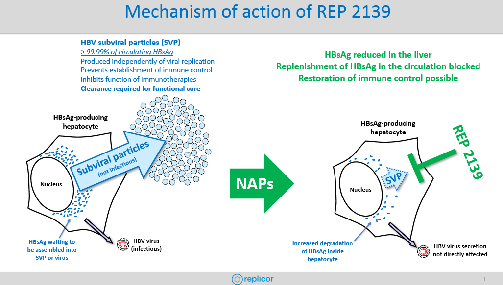
REP 2139 is a polymer built from the building blocks the body uses to store genetic material in the body (nucleic acids). These building blocks are linked together in a unique pattern to form nucleic acid polymers (or NAPs for short) and in the case of REP 2139, use only naturally occurring nucleic acids and modifications to prevent it being recognized as a foreign molecule. As a result, REP 2139 is very well tolerated and safe in clinical trials.

In HBV infection, the most abundant viral antigen in the blood is the hepatitis B surface antigen (HBsAg) which plays an important role in preventing immune control of HBV. Circulating HBsAg is almost entirely in the form of non-infectious HBV subviral particles (SVP) which are produced independently from viral replication, making this viral antigen difficult to target with approved therapies. REP 2139 naturally enters liver cells (hepatocytes) and blocks the assembly of SVP in any hepatocyte producing SVPs. This mechanism effectively blocks the replenishment of HBsAg in the blood and also reduces HBsAg inside these hepatocytes. The overall antiviral effect of REP 2139 is to allow the body to clear HBsAg in order to reduce or remove the inhibition of immune control caused by this viral antigen.

2. REP 2165 is also mentioned as a drug candidate. Can you explain the difference between REP 2139 and REP 2165.

REP 2165 is a close cousin of REP 2139 being examined for potential use in future therapy with more frequent dosing to improve HBsAg response in selected cases and was proven to be as effective as REP 2139 in this study. More information about REP 2165 can be found under question 6.

3. Can you share the latest results from phase 2 trials? How is REP 2139 administered to patients, and for what duration of time? What kind of side effects can patients expect with REP 2139?

In our latest trials, side effects have been limited to mild effects from pegylated interferon (pegIFN). REP 2139 is currently given in a formulation (REP 2139-Mg) which results in little to no side effects during administration. REP 2139 is currently administered once every week for 48 weeks by intravenous infusion in combination with other antiviral agents. REP 2139-Mg is expected to be as effective with a once weekly injection under the skin (subcutaneous injection) which will be used in future trials.

4. Is REP 2139 equally effective in HBeAg positive and negative patients?

REP 2139 is effective in HBeAg positive and in HBeAg negative patients in multiple genotypes. As REP 2139 targets a host protein involved in SVP formation and not the virus or SVP directly, its antiviral effects are expected to be similar in all HBV genotypes and may also be effective in the presence of other co-infections with HBV such as HCV and HIV.

5. Can REP 2139 be safely used in patients with cirrhosis?

Another of the remarkable features of NAP based therapy is the high rate of flares in liver transaminases during therapy (occurring in almost all participants in the REP 401 study). Patients with these flares had no symptoms or any negative impact on their liver function.

Continually expanding evidence in the field tells us that during treatment of HBV, these flares are signs of elimination of HBV infection from the liver and are not accompanied by changes in liver function. These same features appear to hold true for transaminase flares during NAP therapy and, when occurring in the absence of HBsAg in the blood, are highly correlated with functional cure in our clinical trials. The ability of cirrhotic patients to tolerate these flares will be tested in future trials and we are encouraged by recent results (produced by a different group) with pegIFN in HBV / HDV co-infected patients showing that host mediated transaminase flares may also be well tolerated in cirrhotic patients.

6. Do you anticipate combination therapy will be needed? Will combination therapy include immune modulators like pegylated interferon and/or treatment with antivirals?

Replicor’s latest REP 401 study is the first in the field to feature triple combination therapy: Tenofovir disoproxil fumarate (TDF), pegIFN and either REP 2139-Mg or REP 2165-Mg. REP 2165 is a close cousin of REP 2139 being examined for potential use in future therapy with more frequent dosing to improve HBsAg response in selected cases and was proven to be as effective as REP 2139 in this study. In addition to the excellent control of HBV DNA with TDF exposure, this triple combination therapy for 48 weeks led to meaningful HBsAg decline (greater than a 10-fold reduction from baseline) in 90 % of participants, HBsAg clearance to very low levels similar to HBsAg “negative” in the qualitative test used in the United States (< 0.05 IU/mL) and HBsAg seroconversion (often with very high titers of anti-HBs antibodies) in 60% of participants. After removal of all treatment (including TDF), a 48-week follow-up yielded very encouraging results: 89% had normal liver function, 56% had reduced liver inflammation, 39% had stable virologic control and an additional 39% had functional cure with HBsAg seroconversion. These results illustrate the effectiveness of combining potent HBsAg reduction with immunotherapy but also suggest that direct acting antivirals such as TDF and entecavir may also contribute to establishing functional cure in a combination setting.

7. Surface antigen loss is key to people living with chronic HBV. Do you believe REP 2139 can provide a functional cure for chronic HBV?

In an early clinical study using NAPs alone, HBsAg clearance by itself resulted in virologic control (low level infection with normal liver infection no longer requiring therapy under current guidelines ) or functional cure (complete control of HBV DNA and HBsAg) persisting after removal of all therapy only in a small proportion of patients but stable throughout a 5 year follow-up. Importantly, HBsAg clearance with REP 2139 in a subsequent study led not only to a dramatic improvement in the activity of various immunotherapies (including pegIFN) but to virologic control occurring in a larger proportion of patients after removal of therapy persisting throughout more than 2 years of follow-up. As a result of these early studies, Replicor believes that the best approach to achieving functional cure of HBV infection is to simultaneously combine potent HBsAg reduction using REP 2139 with immunotherapy to restore effective and long-lasting immune control.

8. Which countries do you anticipate phase 3 trials to occur? Do you anticipate trials in the U.S?

Replicor believes that the combination of therapy with NUCs such as TDF and ETV, pegIFN and REP 2139-Mg will be the first available therapy to offer patients a real chance of eliminating the need for therapy and establishing functional control of their HBV infection and normalizing their liver function. Work is ongoing to start a phase II US trial in collaboration with the Aids Clinical Trials Group as soon as possible. We are also planning to assess other immunotherapies, the effectiveness of which we believe will be similarly improved with HBsAg clearance as we have demonstrated for pegIFN.

9. With regard to hepatitis delta, is there a difference in the mechanism for how it works?

REP 2139 is also potently active against HDV infection and is able to rapidly eliminate HDV RNA, normalize liver function and reverse the liver inflammation associated with HBV / HDV co-infection. The completed follow-up results from our long term follow-up study of co-infected participants treated with REP 2139 and pegIFN show complete control of HDV infection at 3.5 years follow-up in the absence of all therapy in a large proportion of patients. In many patients this control of HDV infection was associated with functional cure of HBV and in some patients with virologic control of HBV. This potent effect against HDV infection is assumed to be driven not only from the effect of REP 2139 on SVP (which also forms the envelope of the HDV virus) but on the ability of REP 2139 to interact with different forms of the hepatitis delta antigen protein essential for HDV replication and assembly.

Thank you to Dr. Vaillant for taking the time to talk to us about REP 2139. The results look promising! We look forward to learning more from continuing and new trials with REP 2139, used alone and in combination with antivirals and immune modulators. We know the hepatitis B virus is challenging, but those living with chronic HBV look forward to a day when there are therapies resulting in a durable loss of surface antigen and sustained viral suppression in a reasonable, finite amount of time. 

Hepatitis B Research Review – February

Welcome to the Hepatitis B Research Review! This monthly blog shares recent scientific findings with members of Baruch S. Blumberg Institute (BSBI) labs and the hepatitis B (HBV) community. Technical articles concerning HBV, Hepatocellular Carcinoma, and STING protein will be highlighted as well as scientific breakthroughs in cancer, immunology, and virology. For each article, a brief synopsis reporting key points is provided as the BSBI does not enjoy the luxury of a library subscription. The hope is to disseminate relevant articles across our labs and the hep B community. 

This paper from the University of Duisburg-Essen in Germany shows that hepatocytes infected with HBV exhibit innate immune signaling via the pattern precognition receptor (PRR) Toll-Like Receptor 2 (TLR2). The adaptive immune response to HBV infection is well characterized and is broken into phases based on serological testing of antibodies produced against the virus. However, whether HBV infection triggers an innate immune response has remained controversial, with the long-held belief being that HBV evades the innate immune system as a “stealth virus”. Contrary to this view, studies of acute HBV infection in patients have indicated an early, innate immune response to HBV characterized by a natural killer (NK) cell response. Toll-like receptors (TLRs) are a class of membrane-bound receptor proteins which play a key role in innate immunity by recognizing foreign pathogens and activating inflammatory signaling cascades. A previous publication from this group has demonstrated that primary human hepatocytes (PHHs) can be stimulated through the TLR proteins TLR1-9. In this paper, PHHs from human donors were infected with HBV ex vivo. Then, expression of the innate immune cytokines Interleukin 1 Beta (IL1B), Interleukin 6 (IL6), and Tumor Necrosis Factor Alpha (TNFα) were measured by quantitative, reverse-transcription polymerase chain reaction (qRT-PCR). HBV-infected PHHs showed greatly increased expression of these genes at three hours after infection compared to mock-infected and not treated PHHs. Additionally, immunocytochemical staining revealed translocation of the transcription factor nuclear factor kappa-light-chain-enhancer of activated B cells (NFκB) to the nuclei of HBV-infected PHHs, indicating a cytokine response. Next, to characterize the innate immune response caused by HBV infection, a DNA microarray was used. Here, PHHs were either infected with HBV or treated with a known TLR ligand such as Pam3Cys (TLR2 agonist) or poly(I:C) (TLR3 agonist). Then, RNA was extracted from the cells and converted through a complementary DNA (cDNA) intermediate into biotin-labeled anti-sense RNA (aRNA) which was then hybridized to a Human Genome U219 Array Plate. This plate, coated with over 530,000 DNA probes representing over 20,000 human genes served as a scaffold for complementary base-pair binding of the aRNAs derived from the cells. Once bound to the microarray, the biotin-labeled aRNAs were detected by staining with streptavidin phycoerythrin, resulting in a fluorescent signal wherever complementary base-paring occurred. This microarray analysis revealed which specific inflammatory genes were up-regulated in the PHHs by each stimuli. Gene expression signals which were induced by HBV infection were compared with those induced by the TLR agonists. The gene expression profile of HBV-infected PHHs was most similar to that of PHHs treated with the TLR2 agonist Pam3Cys. This data indicates that HBV infection induces a TLR2-like innate immune response. Importantly, no expression of interferon-stimulated genes (ISGs) was detected in the microarray analysis. Finally, PHHs were pre-treated with neutralizing antibodies against TLR2 (nABTLR2) prior to infection with HBV. HBV-mediated induction of IL1B, IL6 and TNF was significantly reduced by nABTLR2 pre-treatment and conversely, HBV replication was increased. In summary, this paper shows that PHHs exhibited an innate immune response to HBV infection via the TLR2 pathway. The group suggests that this response is one of the body’s first steps leading to HBV clearance. Furthermore, in the discussion section the group indicates that the HBV surface antigen (HBsAg) is likely the protein component of HBV which activates TLR2 upon infection. This finding may help in the development of strategies to cure chronic HBV infection.

​This paper from Wuhan University in China reports that HBV infection can increase the expression of Programmed Death Ligand 1 (PD-L1) on the surface of infected hepatocytes, allowing them to escape destruction by the adaptive immune system. PD-L1 is the binding partner of Programmed Death 1 (PD-1), an immune checkpoint protein on the surface of T cells. The expression of PD-L1 on cell surfaces allows for their recognition by circulating T cells as part of the body and not an outside threat. This interaction is important for the prevention of autoimmune disorders in which the immune system attacks healthy cells of the body. However, PD-L1 is commonly over-expressed in a number of cancers and is a hallmark of especially aggressive cancers. PD-L1 expression on cancer cells allows them to neutralize T cells which specifically target them. This is one example of an “immune-escape” strategy exhibited by cancers. Accordingly, PD-L1 and PD-1 are the target of a number of FDA approved immunotherapies for cancer including the PD-L1 inhibitors Tecentriq, Bavencio, and Imfinzi and the PD-1 inhibitors Keytruda, Opdivo, and Libtayo. These drugs are some of the first in their class in that they are not small molecules, but are recombinant, monoclonal antibodies. Phosphatase and tensin homologue deleted on chromosome 10 (PTEN) is a tumor suppressor which is mutated or deleted in many human cancers. PTEN is a phosphatase, a protein which dephosphorylates other molecules. This group has previously shown that PTEN plays a role in antiviral innate immunity. Therefore, they wanted to see if PTEN also regulates the adaptive immune response in the context of HBV infection. First, they used immunohistochemical staining of patient liver tissues to compare the levels of PTEN and PDL-1 in patients chronically infected with HBV vs healthy controls. There was a reduced staining of PTEN and a heightened staining of PD-L1 in chronic HBV tissues compared to controls. The group then found a similar correlation using immunofluorescence, qPCR and Western blotting of HepG2 cells vs HepG2.2.15 (HBV-producing) cells. They also transfected HepG2 cells and infected mice via hydrodynamic injection with an HBV-containing vector (pHBV1.3) or an empty vector control (pUC18) and then performed qPCR and/or Western blotting.  In all systems, HBV infection/production induced a reduction of PTEN and an increase in PD-L1 expression. Then, in order to elucidate this phenomenon further, a PTEN-expressing plasmid was transfected into HepG2.2.15 cells, which resulted in a reduction in PD-L1 mRNA and protein. Conversely, PTEN knockdown in HepG2.2.15 cells resulted in a two-fold increase in PD-L1 mRNA and protein expression. These results show that HBV inhibits PTEN expression which in turn causes up-regulation of PD-L1. Next, the group transfected HepG2 and Huh7 cells with a number of constructs conferring individual HBV proteins. They found that HBV X protein (HBx) and HBV polymerase (HBp) reduced PTEN expression more than any other HBV protein components. Next, the group analyzed how HBV production in hepatocytes affected human T cells grown in co-culture. Jurkat T cells were co-cultured with either HepG2 or HepG2.2.15 cells and then analyzed by flow cytometry. Jurkat T cells grown alongside the HBV-producing HepG2.2.15 cells had a higher incidence of apoptosis, a higher expression of PD-1, and less Interleukin-2 (IL-2) secretion than those grown alongside HepG2 cells. This result indicates that HBV-infected hepatocytes suppress local T cell responses by PD-L1/PD-1 signaling. Finally, the group used a mouse model of HBV infection to show that PTEN over-expression promotes HBV clearance in vivo. This paper shows that PD-L1, a highly studied drug target implicated in the immune-escape of cancers is also up-regulated by HBV infection. Furthermore, the HBV proteins responsible for this up-regulation are HBx and HBp. This finding may help in the development of  immunotherapies to treat chronic HBV infection. Perhaps FDA approved PD-L1 or PD-1 inhibitors may be used in conjunction with interferon alpha treatment or HBV antivirals to boost the immune response against HBV-infected hepatocytes.

This paper from National Tsing Hua University in Hsinchu, Taiwan reports the design and testing of nanoparticles which selectively confer immunogene therapy to hepatocellular carcenoma (HCC) cells. Nanoparticles are very small (1-1000nm) particles which have become an attractive novel drug candidate in recent years. The use of nanoparticles as medicine would enable the customizable delivery of DNA, RNA, or protein payloads to cells. The novel nanoparticles presented here deliver both a small interfering RNA (siRNA) against the Programmed Death Ligand 1 (PD-L1) gene as well as a plasmid DNA (pDNA) encoding the cytokine Interleukin 2 (IL-2). The strategy behind the nanoparticles’ design is to both inhibit an immunosuppressive gene (PD-L1) and up-regulate an immunostimmulatory gene (IL-2) in tumor cells. Delivery of such genes to tumor cells would make them more vulnerable to destruction by circulating cytotoxic T cells (CD8+ T cells). This type of approach is needed, because many advanced tumors create an immunosuppressive tumor micro-environment (TME) rendering many cancer treatments ineffective. The nanoparticles presented here are referred to as tumor-targeted lipid dendrimer-calcium phosphate (TT-LDCP) nanoparticles. The nanoparticles consist of a core of calcium phosphate, thymine-capped polyamidomine (PAMAM) dendrimers, siRNA, and pDNA. This core is coated with an inner lipid called DOPA and outer leaflet lipids DOPC, DOTAP, and DSPE-PEG. The nanoparticle is then tagged with SP94 (SFSIIHTPILPL), a polypeptide which selectively binds to HCC cells but not healthy hepatocytes. Dendrimers are repeatedly-branching molecules which exhibit a sphere-like shape. PAMAMs are the most well-characterized class of dendrimers, consisting of branching amide and amine groups. The calcium phosphate and PAMAM dendrimers in the core of the TT-LDCP nanoparticle promote endosomal escape of the nucleic acid payload. Additionally, this group shows that the PAMAM dendrimers in TT-LDCP nanoparticles also activate the STING pathway. The group showed that STING was activated by treating mouse HCC cells HCA-1 with complete nanoparticles or those lacking the dendrimers. Cells treated with complete nanoparticles showed, by Western blot a higher level of both TBK1 and IRF3 phosphorylation than those treated with incomplete nanoparticles. Those cells treated with complete nanoparticles also displayed heightened transcription of the STING-triggered proinflammatory genes Ifnb,Ccl5, and Cxcl10 as measured by qPCR. Furthermore, the group showed that treatment using their nanoparticles of mice bearing orthotopic HCC implants resulted in dendritic cell maturation in those animals, regardless of the identity of the genes delivered. These results indicate that the dendrimers used in the TT-LDCP nanoparticles not only serve for efficient delivery of nucleic acids, but also as adjuvants that stimulate the STING pathway and activate tumor-infiltrating dendritic cells. This publication gives a glimpse into what future therapies for cancer may look like. The nanoparticle designed by this group is unique in that it has multiple functionalities: selectively targeting HCC cells, inhibiting PD-L1 expression, inducing IL-2 expression, and activating the STING pathway. Such a complex design is bound to require fine tuning before it can become a medicine. But a multi-target immunotherapeutic such as this may be exactly what is needed to help the body fight against aggressive, immunosupressive tumors.

Lay Summary: 
This month, the innate immune system was the focus of HBV research. Scientists hope to find how the innate immune system interacts with HBV during viral infection and proliferation. Doing so will shed light on host factors which lead to chronic infection and inform antiviral strategies. Notably, this month a human protein, MX2 was found to have potent anti-HBV activity by preventing cccDNA formation. Also, a microRNA encoded by HBV called HBV-miR-3 was found to activate the human innate immune system to limit HBV replication. This month, a paper studying woodchuck hepatitis virus (WHV) traked activation of the innate immune system as well as he adaptive immune system in an acute infection model. Also this month, concerning hepatocellular carcenoma (HCC), the alternative splicing of mRNA in tumors was found to vary in HCC patients based upon their risk factor (HBV, HCV, or alcohol). Finally, a review was published this month concerning STING, an innate immune protein which is not activated by HBV infection but which may prove a valuable tool for cancer treatment.  

Meet our guest blogger, David Schad, B.Sc., Junior Research Fellow at the Baruch S. Blumberg Institute studying programmed cell death such as apoptosis and necroptosis in the context of hepatitis B infection under the direction of PI Dr. Roshan Thapa. David also mentors high school students from local area schools as part of an after-school program in the new teaching lab at the PA Biotech Center. His passion is learning, teaching and collaborating with others to conduct research to better understand nature.

Hep B Patient Engagement Survey: Help Guide The Future of Hepatitis B Therapies

If you’ve ever wanted to help guide the future of hepatitis B treatments, now is your chance! The Hepatitis B Foundation has created a short survey that is designed to capture a comprehensive view of the patient experience. The survey, which takes approximately 20-25 minutes to complete, will be made available for use by the US Food and Drug Administration (FDA) and drug development researchers to help clinical trial development for future hepatitis B therapies. All survey responses are anonymous.  

All individuals living with chronic hepatitis B are welcome to take this survey! After answering a few standard questions, participants will be asked whether or not they are currently on treatment for hepatitis B. If they are on treatment, the survey will prompt the participant to answer a few questions about their experience with the medication, such as how it makes you feel to take it, 

and what challenges you may face while taking your medication. All participants, regardless of their current treatment status, will have an opportunity to provide feedback on what they hope future medications will look like! 

The patient perspective is essential to creating a treatment that is not only effective but something that those living with hepatitis B would be willing to take. Oftentimes, researchers do not have the opportunity to gather insight into what patients are looking for or how a therapy would impact their lives. This can result in missed dosages of a medication, or avoiding a therapy altogether, rendering the treatment ineffective. This survey is also unique because it seeks to capture the diverse experiences of global patients living with hepatitis B. As we want to ensure that new treatments are as universal as possible, gathering the thoughts of both international and national individuals will ensure that different voices and opinions are captured! 

The survey is the first part of a multistep process to collect information on the patient experience. In the upcoming months, we will carry out focus groups and interviews to have a better understanding of what it is like to live with hepatitis B, its impact on a person’s daily life, and more. The information collected will help to direct our patient-focused drug development meeting – a chance for stakeholders and those living with hepatitis B to meet and discuss the patient perspective –  in June 2020.

The work being done will result in a broader understanding of how individuals are affected by the disease and more informed decisions regarding future hepatitis B therapies. Help guide the future of hepatitis B clinical trials and drug development by taking the survey today! 

Eiger BioPharmaceuticals: Developing Two New Hepatitis Delta Treatments

Eiger is currently working on two new drugs for hepatitis delta; Lonafarnib and Pegylated Interferon Lambda, which are both currently inphase 3 clinical trials. Lonafarnib is a new type of treatment that attempts to control hepatitis delta through a new method: through blocking a key enzyme that is needed for the hepatitis delta virus to replicate. Blocking this enzyme prevents a new virus from being created, which may control and even cure hepatitis delta.

Lambda is being developed as a better tolerated interferon compared to interferon alfa (IFN alfa). Interferons work by stimulating the body’s own immune system to fight the virus. Pegylated interferon alpha, which is the only current treatment for hepatitis delta, is a difficult treatment to tolerate, with many patients experiencing unpleasant side-effects. Lambda utilizes the same method of treatment as IFN alfa, in combination with a new strategy, which stimulates an immune response and targets receptors in the liver, which may reduce side effects and result in improved tolerability.

Below is Eiger Biopharmaceuticals’ response to a series of questions we posed to them. 

Image courtesy of Praisaeng, at FreeDigitalPhotos.net.

1. Lambda is an immunomodulator and Lonafarnib is a prenylation inhibitor. Can you explain in laymen’s terms the mechanism for these drugs and how they work?

Eiger’s wording: Lonafarnib is a well-characterized, first-in-class, orally active inhibitor of an enzyme that is key to a vital process in the life cycle of HDV. Inhibiting this enzyme blocks the ability of HDV to assemble and package viral particles. Currently approved nucleos(t)ide treatments for HBV only suppress HBV DNA, do not affect HBsAg, and have no impact on HDV infection.

Lambda is being developed as a better tolerated interferon compared to interferon alfa (IFN alfa). Lambda is a well-characterized, first-in-class, type III interferon (IFN) that stimulates immune responses that are critical for the development of host protection during viral infections. By targeting receptors that are localized in the liver, Lambda treatment may reduce side effects and result in improved tolerability .

Can you share, in simple terms, the basic results of Eiger phase 3 studies for hepatitis delta trials? Are these drugs equally effective in HBeAg positive and negative HBV patients?

The Eiger Phase 2 LOWR program with Lonafarnib has been completed. Over 120 patients were dosed in Phase 2 dose-finding studies to identify combination regimens of lonafarnib (LNF) and ritonavir (RTV) with and without pegylated interferon-alfa (PEG IFN α), with efficacy and tolerability to enable viral load suppression of HDV RNA and ALT normalization at Week 24.

  • Dosing regimens of LNF 50 mg twice daily + RTV 100 mg twice daily with and without PEG IFN-a-2a 180 mcg once weekly were identified with the following reported results:
    • All-oral: Lonafarnib boosted with ritonavir
    •  29% of patients achieved ≥ 2 log decline and ALT normalization
  •  Combination: Lonafarnib boosted with ritonavir + PEG IFN-a-2a
    •  63% of patients achieved ≥ 2 log decline and ALT normalization

These dosing arms are being further studied in the global Phase 3 D-LIVR study. Phase 2 studies have not been stratified by HBeAg status.

The D-LIVR Study, a Phase 3 pivotal trial, is on-going and evaluating the safety and efficacy of lonafarnib treatments in patients chronically infected with Hepatitis Delta Virus (HDV). Topline Week 48 data will be available in 2021.

2. How will Lambda and Lonafarnib be administered to patients?

Lonafarnib capsules are administered orally twice daily by mouth. Lonafarnib is taken in combination with ritonavir, a therapeutic booster that increases the bioavailability of lonafarnib. Ritonavir tablets are administered orally twice daily by mouth.

Pegylated interferon-lambda is administered as a self-administered subcutaneous injection once weekly.

3. Do you anticipate combination therapy will be needed and if so, which combinations do you anticipate?

No form of viral hepatitis has been cured with a single drug. Combinations of treatments with different mechanism of actions have always been required.

Lonafarnib and interferons have different mechanisms of action and have been studied as monotherapies and in combination together as treatments for HDV. While each treatment alone reduces the HDV viral load, combination studies have shown that using these treatments together leads to a synergistic effect and further reduces the HDV viral load.

Recently, the interim end of treatment results of peginterferon lambda (Lambda) and lonafarnib combination study in HDV-infected patients were presented at AASLD 2019. The LIFT study is being conducted within the National Institutes of Health (NIH) at the National Institute of Diabetes and Digestive and Kidney Diseases (NIDDK). LIFT is a Phase 2a open-label study of 26 adult patients with chronic HDV treated with Lambda 180 mcg once weekly in combination with Lonafarnib 50 mg twice daily boosted with ritonavir 100 mg twice daily for 24 weeks. Primary efficacy endpoint is > 2 log HDV RNA decline at end of treatment. At the time of analysis, 19 of 26 patients had reached Week 24. Median HDV RNA decline was 3.4 log IU/mL (IQR: 2.9-4.5, p<0.0001) with 53% (10 of 19) patients achieving below the limit of quantification and 37% (7 of 19) patients achieving undetectable HDV RNA at Week 24. 18 of 19 patients (95%) achieved primary endpoint of > 2 log decline during 24 weeks of therapy. We believe these data are the most encouraging yet in pursuit of HDV cure.

4. What kind of side effects can patients expect with Lambda and Lonafarnib, with or without combination therapy?

The most common side effects of lonafarnib include diarrhea, nausea, fatigue, decreased appetite, vomiting, abdominal pain, and decreased weight. Antacid, antiemetic, and antidiarrheal medications may be used prophylactically to treat these gastrointestinal side effects.

The most common side effects of pegylated interferon-lambda (Lambda) are the expected side effects of interferons. However, these side effects have been demonstrated to be much milder and less severe than what has been previously been shown with pegylated interferon-alfa (alfa). These include musculoskeletal (myalgia, arthralgia, and back pain), flu-like symptoms (chills, pyrexia, and pain) and elevated alanine aminotransferase (ALT) levels.

A combination of these side effects is expected with combination therapy.

5. Are Lambda and Lonafarnib safe for use in people with cirrhosis?

Currently, the safety and efficacy of lonafarnib and pegylated interferon-lambda are being investigated in persons chronically infected with HDV. The clinical trials require study participants meet certain eligibility criteria to be included in these studies. These eligibility criteria may or may not reflect the type of patient who will use these therapies after they receive FDA approval.

Phase 2 and Phase 3 studies both include patients with well-compensated cirrhosis.

6. With a clearance of HDV, would you also anticipate a loss of surface antigen – functional cure for chronic HBV as well? If so, in what percentage of HBeAg and HBeAb patients?

HDV is always found as a co-infection with HBV because HDV requires just a small amount of HBV surface antigen (HBsAg) to complete HDV viron replication. However, an HDV / HBV coinfection leads to much more severe chronic viral hepatitis compared to HBV monoinfection alone. Therefore, it is important to treat HDV, even if HBV is not cured. It is possible to clear HDV RNA without loss of HBsAg.

7. Lambda and Lonafarnib are currently in phase 3 trials for delta. Are you able to provide an approximate timeline for when it will be approved for use in U.S. and Europe?

Eiger BioPharmaceuticals is committed to developing safe and effective therapies for HDV and providing patients with a pathway to gain access to approved therapies as quickly as possible.

The D-LIVR Study is a global study that is evaluating the safety and efficacy of lonafarnib treatment in patients chronically infected with HDV. The D-LIVR Study is recruiting subjects in up to 20 countries in over 100 study sites. The D-LIVR study includes 48 weeks of treatment with two different lonafarnib-based treatment regimens, followed by 24 weeks of follow-up. Primary endpoint is ≥ 2 log decline and ALT normalization at Week 48. Topline data from the Phase 3 D-LIVR study will be available in 2021. For more information about study locations and eligibility, please visit www.clinicaltrials.gov  (NCT03719313).

End of Phase 2 meeting with FDA to discuss Phase 3 development with Lambda monotherapy is planned for Q1 2020.

Hepatitis B Research Review


Welcome to the Hepatitis B Research Review! This monthly blog shares recent scientific findings with members of Baruch S. Blumberg Institute (BSBI) labs and the hepatitis B (HBV) community. Technical articles concerning HBV, Hepatocellular Carcinoma, and STING protein will be highlighted as well as scientific breakthroughs in cancer, immunology, and virology. For each article, a brief synopsis reporting key points is provided as the BSBI does not enjoy the luxury of a library subscription. The hope is to disseminate relevant articles across our labs and the hep B community. 

Interferon-inducible MX2 is a host restriction factor of hepatitis B virus replication Journal of Hepatology

  • This paper from Fudan University in Shanghai, China reports the interferon-induced GTPase MX2 as a host protein which inhibits HBV replication. Interferon alpha (IFN-α) is a type 1 interferon used in a subset of HBV-infected patients to help eradicate the virus. IFN-α treatment results in the activation of hundreds of genes known as interferon-stimulated genes (ISGs). Which ISGs are most important in eliminating HBV infection remain largely unknown. GTPases are a large family of hydrolase enzymes which convert guanosine triphosphate (GTP) to guanosine diphosphate (GDP). GTPases act as molecular switches in an array of cellular process including signal transduction, cell division and differentiation, and protein translocation. The myxovirus resistance (Mx) proteins are highly conserved, dynamin-like, large GTPases. Humans have two MX proteins: MX1 and MX2, both of which are known ISGs. While MX1 is known to have broad-spectrum antiviral activity against RNA viruses, MX2 has only recently been shown to inhibit human immunodeficiency virus 1 (HIV-1), hepatitis C virus (HCV), and hepesviruses. MX2 antiviral activity against HIV-1 and herpesviruses is mediated through MX2 binding to the capsid of invading viruses whereby it likely inhibits the uncoating of viral DNA. In HCV, MX2 was found to interact with non-structural protein 5A (NS5A) thereby inhibiting its localization to the endoplasmic reticulum (ER). MX1 has been reported to inhibit HBV replication by inhibiting nuclear export of viral RNas and/or trapping the HBV core protein indirectly. This study investigates the anti-HBV activity of MX2. First, the group compared the anti-HBV activity of MX2 to four other innate immune restriction factors: HNRNPU, SAMHD1, MOV10 and A3G. They co-transfected these genes along with the HBV genome into HUH-7 cells and then assessed HBV replication via Southern blot. MX2 was found to inhibit HBV replication the most, with 44% of viral DNA compared to the empty vector control. The group then used siRNA, Southern blot, Western blot, fractionation, and mutagenesis studies to elucidate the anti-HBV role of MX2. Overall, they found that MX2 significantly reduces HBV RNA levels and indirectly impairs cccDNA formation. MX2 was found to contribute substantially to the anti-HBV affect of  IFN-α. Both the GTPase activity and oligomerization status of MX2 were found to be important in conferring its anti-HBV affect. In the future, MX2 and its related pathways may be exploited to help prevent the formation of and even eliminate cccDNA in those infected with HBV.

An HBV-encoded miRNA activates innate immunity to restrict HBV replication – Journal of Molecular Cell Biology

    • This paper from the Tianjin Medical University in China explains how an HBV-encoded microRNA (miRNA) activates the innate immune system in humans infected with the virus. miRNAs are short (21-25 nucleotides) sequences of mRNA which are mainly involved in post-transcriptional silencing of genes. miRNAs are produced in plants, animals, bacteria, and viruses. Typically, miRNA acts to silence protein translation from a messenger RNA (mRNA) by binding to the 3′ untranslated region (UTR) of the mRNA. This binding may result in the destabilization or cleavage of the mRNA or inhibit the function of the ribosome during translation. This group has identified an miRNA from the HBV genome called HBV-miR-3 which they have previously reported inhibits HBV replication by targeting the HBV mRNA transcript. In this paper, the group first shows that HBV-miR-3 is produced in an amount proportional to virus infection in vitro. They also show that HBV-miR-3 is secreted from cells in exosomes. Next, using both patient serum samples and in vitro assays, the group found a positive correlation between HBV-miR-3 production and IFN-α signaling pathways. In patient serum, levels of HBV-miR-3 positively correlated with levels of the hepatitis-related parameters alanine aminotransferase (ALT), aspartate transaminase (AST) and type I IFNs (IFN-α and IFN-β). In cell culture, they observed an increased expression of  the IFN-α-induced antiviral effectors OAS-1, MX1, IFIT2 and IFIT3 in the context of HBV-miR-3 production. Further experiments indicated that HBV-miR-3 promotes IFN-α production by suppressing the expression of suppressor of cytokine signaling 5 (SOCS5), allowing for signal transducer and activator of transcription 1 (STAT1) to be activated by phosphorylation. Finally, the group shows that HBV-miR-3 released from infected cells in exosomes  promotes polarization of the M1 macrophage phenotype. M1 or “classically activated” macrophages secrete high levels of pro-inflammatory cytokines and thereby fight pathogenic infections. Taken together, these results show that aside from directly limiting HBV replication, HBV-miR-3 also indirectly limits HBV infection by activating the host innate immune system. The virus may do this in order to adopt host miRNA-mediated antiviral machinery and thereby alleviate pathogenesis so that persistent and latent infection can continue. In the future, levels of HBV-miR-3 may be used as a diagnostic marker for HBV infection and may shed light on novel antiviral approaches.

Innate and adaptive immunity associated with resolution of acute woodchuck hepatitis virus infection in adult woodchucks – PLOS Pathogens

    • This paper from Georgetown University in Washington, DC is a “woodchuck paper”. That is, it is an in vivo study of woodchucks infected with Woodchuck Hepatitis Virus (WHV). WHV infection is used as a model system for HBV infection in humans because WHV is similar to HBV. This type of study is beneficial, especially when studying the immune response to hepadnaviruses, because humans infected with HBV are typically asymptomatic in the early stage of infection and because it is not advisable to obtain liver biopsies from these patients. The woodchuck infection model offers a controlled infection with WHV at a known time-point, which can be monitored by regular blood tests and liver biopsies. When studying the immune response to hepadnaviruses, liver biopsies are necessary because the liver is the site of the infection. About 95% of adults infected with HBV “clear” the virus; that is, their immune system is able to fight off the virus completely, giving them life-long immunity. The other 5% become chronic carriers of HBV and are at a high risk for liver cirrhosis and hepatocellular carcinoma (HCC). However, 95% of infants infected with HBV become chronic carriers. Differences in the immune systems of adults vs infants have been attributed to this drastic difference in chronicity, but what specific components of the immune system are important in staving off chronic infection remain unknown. Overall, the data presented here indicate that there is an early, non-cytolytic control of WHV replication mediated by interferon gamma (IFN-γ) produced mainly by natural killer (NK) cells. This was followed by an adaptive immune response characterized by antibody production, a T-cell response, and cytolytic action of cytotoxic T lymphocytes (CTLs). This adaptive immune response led to both the decline of WHV as well as symptoms of acute hepatitis B (AHB) including sinusoidal and portal inflammation in the liver.

Differential alternative splicing regulation among hepatocellular carcinoma with different risk factors BMC Medical Genomics

    • This paper from the University of Utah School of Medicine in Salt Lake City, Utah uses bioinformatics to examine how different risk factors for hepatocellular carcinoma (HCC) correlate with differential alternative splicing (AS) of tumor mRNAs. After a primary (precursor) mRNA transcript is produced in the nucleus by RNA polymerase, the transcript must “mature” by having regions called “exons” removed in a process called splicing. Splicing results in an mRNA transcript consisting entirely of “introns”. The mRNA is then capped at its 5′ end with a 7-methylguanosine residue and polyadenylated at its 3′ end with about 200 adenylate residues (poly-A tail). This mature mRNA is able to exit the nucleus and be translated into protein by a ribosome. Alternative splicing (AS) describes how one genomic region may code for many different protein variants (isoforms) by differential spicing of the primary mRNA transcript. A common mechanism of AS is “exon skipping”, where exons are included in some mature transcripts but not others. HCC has various risk factors including alchohol consumption and infection with hepatitis B or C viruses (HBV and HCV). This study used data from The Cancer Genome Atlas (TCGA) and  the Genomic Data Commons (GDC) Data portal to analyze 218 patients with primary HCC associated with HBV (n = 95), HCV (n =47), or alcohol (n = 76). They used RNA sequencing (RNA-Seq) data to examine differences in AS between three groups: HBV vs. HCV, HBV vs. alcohol, and HCV vs. alcohol. 143 genes were identified with differential AS across these groups and these genes were found to be mainly involved in immune system, mRNA splicing-major pathway, and nonsense-mediated decay pathways.Of the 143 AS genes identified, eight and one gene were alternatively spliced specific to HBV and HCV respectively. The human leukocyte antigen genes HLA-A and HLA-C had differential AS in HBV-related HCC compared to both HCV- and alchohol-related HCC. HLA ptoteins are part of the major histocompatibility complex (MHC) class 1 surface proteins which present foreign antigens to the immune system. Also, exon 3 of  the gene encoding inositol hexakisphosphate kinase 2 (IP6K2) was skipped more often in HBV-related HCC than in other groups. IP6K2 is known to be involved in cancer metastasis. This study represents the first investigation into how different risk factors of HCC may affect the AS status of specific genes.

The Cytosolic DNA-Sensing cGAS–STING Pathway in Cancer (Review) Cancer Discovery

    • This review from the Memorial Sloan Kettering Cancer Center in New York City covers current understanding of the cGAS-STING pathway in the context of cancer. While it is well known that the cGAS-STING pathway is an evolutionarily-conserved  antiviral signaling platform, how this pathway is involved in tumorigenesis remains unclear. In preneoplastic (early tumor) cells, cGAMP produced in response to DNA damage is exported out of the cell to activate STING in neighboring antigen-presenting cells (APC). This activation results in the release of type 1 interferon (IFN) from the APC, which cross-primes natural-killer and CD8 T-cells to kill the preneoplastic cells. In this context, the cGAS-STING pathway plays a role in tumor surveillance by activating innate immunity to create “hot spots” of inflammation. However, there is also evidence that activation of the cGAS-STING pathway can contribute to tumorigenesis.  In advanced, metastatic tumor cells, chronic activation of STING by chromosomal abnormalities leads to suppressed production of IFN and the upregulation of Nf-kB-driven pro-survival genes. This can drive chronic inflammation of the tumor as well as its metastasis to other locations in the body. Activation of the STING pathway in tumor cells may also allow for their immune evasion by inducing autophagy and upregulating expression of programmed death-ligand 1 (PD-L1). Another interesting finding mentioned in this review is a STING-independent form of cGAS activation which may drive tumorigenesis during cell division. During mitosis, cytoplasmic cGAS may bind to repeat sequences in the centromere regions of chromosomal DNA. Once bound, cGAS may interrupt the repair of sister chromatids by homologous recombination, causing aneuploidy in daughter cells, a hallmark of tumor cells. Of additional interest, mentioned in this review are several recent findings regarding the cGAS-STING pathway, including: cGAS can be activated by extracellular DNA entering the cell in exosomes; cGAS can be activated by “micronuclei” which are small nuclear compartments in the cytoplasm formed by chromosomal instability; cGAS-DNA complexes turn into a liquid phase to produce cGAMP; STING dimers oligomerize to form tetramers when activated; palmitoylation of STING has been proposed to recruit TANK binding kinase 1 (TBK1) and interferon regulatory factor 3 (IRF3).

Lay Summary: 
This month, the innate immune system was the focus of HBV research. Scientists hope to find how the innate immune system interacts with HBV during viral infection and proliferation. Doing so will shed light on host factors which lead to chronic infection and inform antiviral strategies. Notably, this month a human protein, MX2 was found to have potent anti-HBV activity by preventing cccDNA formation. Also, a microRNA encoded by HBV called HBV-miR-3 was found to activate the human innate immune system to limit HBV replication. This month, a paper studying woodchuck hepatitis virus (WHV) traked activation of the innate immune system as well as he adaptive immune system in an acute infection model. Also this month, concerning hepatocellular carcenoma (HCC), the alternative splicing of mRNA in tumors was found to vary in HCC patients based upon their risk factor (HBV, HCV, or alcohol). Finally, a review was published this month concerning STING, an innate immune protein which is not activated by HBV infection but which may prove a valuable tool for cancer treatment.  

Meet our guest blogger, David Schad, B.Sc., Junior Research Fellow at the Baruch S. Blumberg Institute studying programmed cell death such as apoptosis and necroptosis in the context of hepatitis B infection under the direction of PI Dr. Roshan Thapa. David also mentors high school students from local area schools as part of an after-school program in the new teaching lab at the PA Biotech Center. His passion is learning, teaching and collaborating with others to conduct research to better understand nature.