Hep B Blog

Category Archives: Hepatitis B Research

If You Have Hepatitis B, Donating Your Blood May Change the Face of Hepatitis B Testing.

The Hepatitis B Foundation has partnered with Plasma Services Group to educate people living with Hepatitis B about the critical need for blood donation. This is not like the local blood drives you always hear about. Instead, Plasma Services Group focuses on specialty plasma collection which supports the making of diagnostic tests used in labs around the world. The demand for HBV testing grows every year, but access to those tests is not assured. As you know, only 25% of people in the U.S. and 10% of people worldwide with Hepatitis B have been diagnosed. With your help, we can reduce those real-life barriers to Hepatitis B testing and improve lives. Follow the link.

How do I donate?

Donating your blood to Plasma Services Group is easy. After you complete this form, they will reach out to you if you are a good candidate for blood donation. If chosen, they will send a phlebotomist to your home to complete the blood-draw.  PSG compensates participants financially as a thank you for the trust, time and efforts associated with donation. This program is only available to U.S. residents who are preferably in the Northeast. You must be 18 years of age or older and weight 110 pounds or more. You must be living with chronic Hepatitis B, which means you have had Hepatitis B for over 6 months.

Why this is important to the future of Hepatitis B?

As you may know, access to good healthcare isn’t always easy. By creating new blood tests, we can help diagnose Hepatitis B more reliably which helps more people get into care and manage their hepatitis B. Your blood donation could directly impact the detection, care and quality of life for millions of people living with hepatitis B who have not been diagnosed yet, as well as those who are managing their care on a daily basis.

Despite the large population of people living with hepatitis B, it is hard for companies that source biological raw materials to recruit donors. Most people are unaware of the large amount of blood plasmas that are essential to manufacture test kits. Rarer subtypes that are prevalent in Africa and Asia, where the need for detection is the highest and growing the fastest, are even harder to find in N. America. By becoming a regular donor to Plasma Services Group, you are filling a vital role for the medical diagnostic industry and helping to close the gap between patient and care.

Get started today!

Fill out this form and Plasma Services Group will fill you in on next steps.

Reactivation with Hepatitis B: Understanding Risk Factors and Prevention Strategies

Understanding the hepatitis B virus and the panel of blood tests needed to determine infection or immunity can be a stressful and challenging task. In simplest terms, “hepatitis” means liver inflammation and the hepatitis B virus can ultimately cause liver inflammation. The liver is an important organ in the human body and responsible for the removal of toxins and regulation of digestion (learn more about the function of the liver here). The hepatitis B virus can infect and disrupt critical functions of the liver in supporting your overall health. 

How the hepatitis B virus works 

In the case of the hepatitis B virus, the host is the liver cell. As the virus makes more copies of itself, the liver may become damaged, and sometimes it is unable to carry out its essential tasks to regulate metabolism, nutrients, and digestion. It is best to prevent hepatitis B infections when we can – and since antibodies are the best defense against the virus, the hepatitis B vaccine can be used to signals the body to make antibodies to fight the virus. The hepatitis B vaccine provides lifelong protection from the virus. However, this is only possible before infection with the virus. If somebody is already infected with the virus, antiviral therapy is used to control the virus and prevent liver damage – antiviral medications disrupt the life cycle of the virus by disabling viral receptors from binding to liver cells. 

Blood test panel to diagnose hepatitis B: 

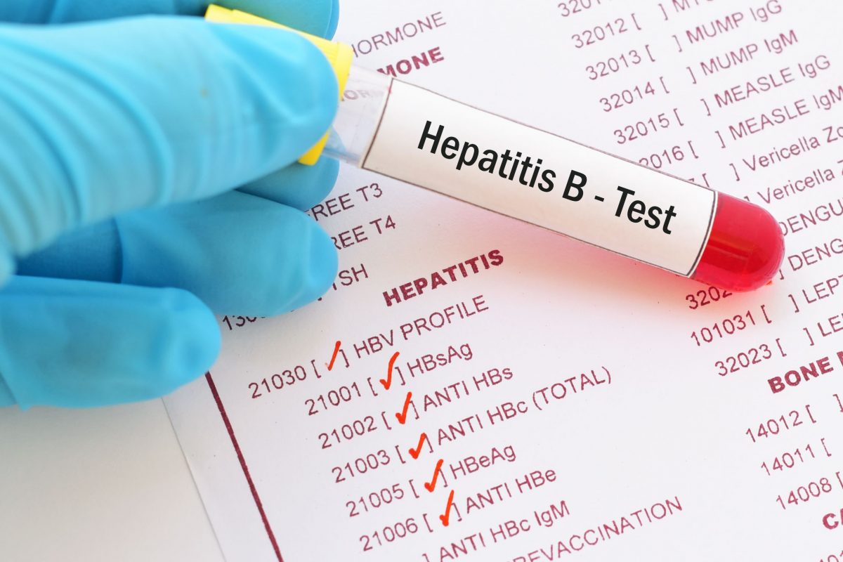
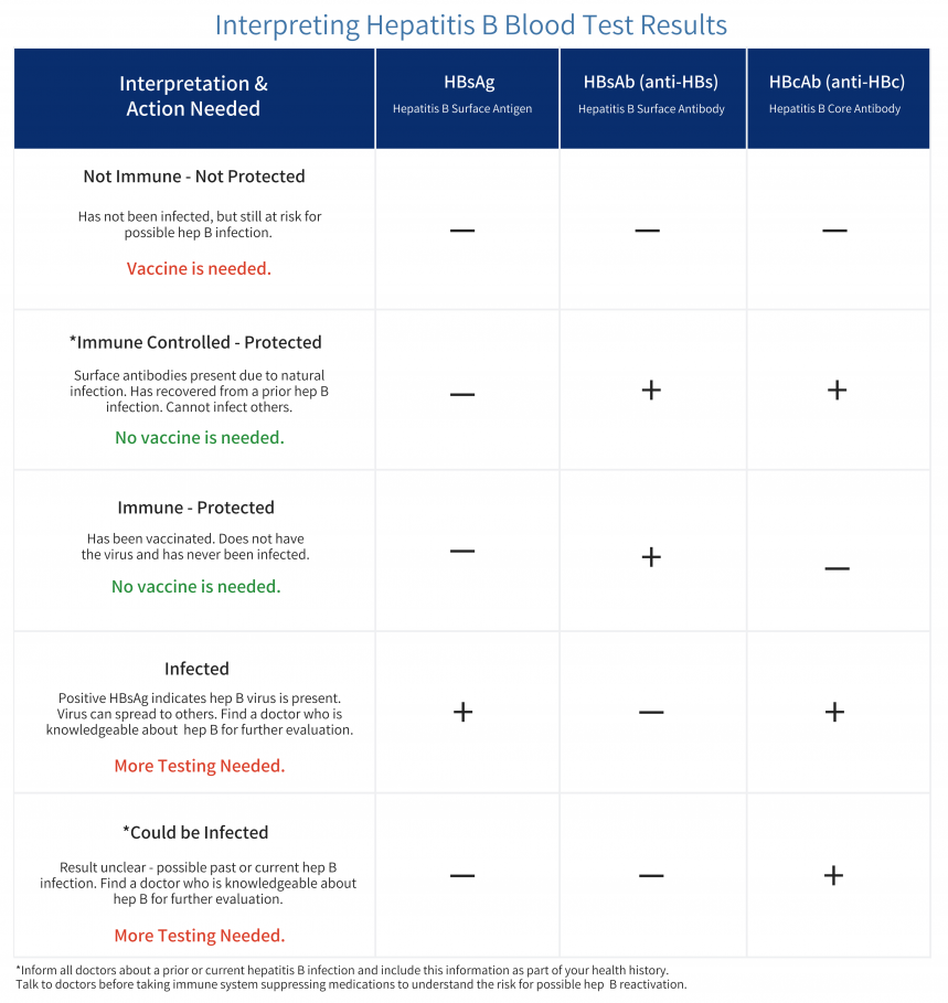
The only way to tell someone’s hepatitis B status is through a panel of blood tests – the tests are all done at one time, and only one small tube of blood is needed. These tests are not included in routine testing, so it is important to ask your doctor to test you for hepatitis B or try to find a free screening event near you (http://www.hepbunited.org/). The panel consists of the following tests to determine your hepatitis B status: 

  1. HBsAg: 
    • This tests for the hepatitis B surface antigen in someone’s blood. The surface antigen is the protein that surrounds the virus and protects it from attack by the host. A positive surface antigen test indicates that the virus is present in the body. A “positive” or “reactive” result for HBsAg indicates that someone is infected with hepatitis B and can transmit the virus to others.  
  1. HBsAb 
    • This tests for the hepatitis B surface antibody in someone’s blood. The surface antibodies are produced by the immune system and can fight off the virus by attaching to the surface antigen protein. This test can detect the presence of these antibodies. Ideally this test will be ordered quantitatively (numerically). A “positive” surface antibody test (meaning numbers reading >10 IU/mL) means that a person has protection against the hepatitis B virus (either by vaccine or from a past exposure).  
  1. HBcAb (total) 
    • This is known as the hepatitis B core antibody test. The core antibody is produced by the immune system after infection with the virus. This test indicates an existing or past infection of the hepatitis B virus.  


To learn more about interpreting your test results, click here. 

Important things to know about Hepatitis B Core Antibody (HBcAb) 

Someone who has markers of past infection, particularly hepatitis B core antibody, can be at risk for hepatitis B reactivation. Reactivation can be triggered by immunosuppressive therapies and cause significant life-threatening challenges. If you test HBcAb+, please talk to your doctor about what that means, and make sure you notify all future health care providers. 

How is reactivation with HBV defined? 

Reactivation is defined as the sudden increase or reappearance of HBV (hepatitis B virus) DNA. When the virus invades the cell, it forms a covalently closed circular DNA (cccDNA) in the nucleus of infected cells referred to as hepatocytes. Because cccDNA is resistant to antiviral treatments, it is never removed from the cells. Therefore, even after recovery from a past infection, the cccDNA is present and may reactivate. It is not clearly understood why this may happen, but certain factors may increase the risk for reactivation.  

To learn more about the core, click here. 

What puts one at risk for reactivation? 

  1. Virologic factors such as high baseline HBV DNA, hepatitis B envelope antigen positivity (HBeAg), and chronic hepatitis B infection that persists for more than 6 months.
    • Detectable HBV DNA levels and detectable levels of HBsAG can increase the risk for HBRr (reactivation) 
    • Testing positive for HBeAg also increases the risk for reactivation 
  2. Co-infection with other viruses such as hepatitis C or hepatitis Delta 
  3. Older age 
  4. Male sex 
  5. Cirrhosis 
  6. An underlying condition requiring immunosuppressive therapies (rheumatoid arthritis, lymphoma, or solid tumors) 
    • Certain medications can increase the likelihood of reactivation by more than 10%.  
    • B-cell depleting agents such as rituximab, ofatumumab, doxorubicin, epirubicin, moderate or high-dose corticosteroid therapy lasting more than 4 weeks. 

How to prevent reactivation of hepatitis B 

Hepatitis B reactivation is a serious condition that can lead to health complications, Reactivation is avoidable if at-risk individuals are identified through screening. Current guidelines recommend that individuals at the highest risk (those receiving B-cell depleting therapies and cytotoxic regimens) should receive antiviral therapies as prophylaxis before beginning immunosuppressive therapy. These antiviral therapies should also be continued well beyond stopping the immunosuppressive therapies. Be sure to talk to your doctor to be sure you are not at risk for reactivation.  

References 

Hepatitis b virus reactivation: Risk factors and current management strategies.

Reactivation of hepatitis B virus: A review of Clinical Guidelines.

https://aasldpubs.onlinelibrary.wiley.com/doi/10.1002/cld.883

https://www.hepb.org/prevention-and-diagnosis/diagnosis/understanding-your-test-results/

GlaxoSmithKline Recruiting for B-Together Hep B Clinical Trials

The company GlaxoSmithKline (GSK) is launching a new clinical trial, called B-Together, that will investigate how two study drugs might work together to treat chronic hepatitis B (CHB). Researchers are hoping to find new potential treatments that could be more effective than those that are currently available and could lead to positive results that last long after the treatment ends. Participants in this trial could play a role in shaping science and changing the landscape of CHB treatment around the world, and will have an opportunity to learn more about the disease itself.

The two drugs that will be investigated in this trial are GSK3228836 and pegylated interferon, also known as Pegasys. In a previous Phase 2 trial, people living with CHB received GSK3228836 for 4 weeks. The Phase 2b B-Together trial will test longer treatment with GSK3228836, followed by Pegasys, to see what effects this may have on viral antigens (such as HBsAg) in the body. 

About the Study Drugs

GSK3228836 is an investigational drug being tested as a potential treatment for CHB, meaning it is not yet approved for this purpose. Current medicines available to treat CHB only stop the virus from multiplying – they do not enable the body to fully clear the infection, so people have to keep taking these medicines. GSK3228836 is designed to stop the virus from producing proteins that may prevent the immune system from fighting the virus. Thus, the study drug may potentially allow the body to gain control over the infection.

The other drug used in this study, Pegasys, is a medicine that is already used on its own by doctors to treat CHB. Pegasys works by enhancing the body’s immune response to viral infections such as hepatitis B.

What Will Happen During This Trial?

During this trial, all participants will receive GSK3228836 followed by Pegasys. After you have finished treatment with GSK3228836, your doctor will check if it is appropriate for you to start treatment with Pegasys. If it is not appropriate, you may not receive Pegasys at all. At the beginning of the trial, you will be assigned by chance to one of two groups. Each group will receive the study drugs for different lengths of time. You will know which group you are in. The B-Together trial lasts about 79 weeks for each participant. This includes a screening period, a study treatment period, and a follow-up period.

Screening Period

At a screening visit, the study doctor will give you a physical examination, ask about your medical history, and conduct medical tests. The screening period may last up to about 6.5 weeks while the study doctor reviews the results of your screening visit to determine if you meet all requirements for participation.

Trial Treatment period

While receiving GSK3228836, you will visit the clinic for either 12 or 24 weeks. For the first two weeks of your treatment with GSK3228836, you will visit twice per week and for the remaining weeks you will visit the clinic once per week.

When you have finished treatment with GSK3228836, your doctor will assess if it is appropriate for you to start treatment with Pegasys. If it is appropriate, then you will then receive treatment  with Pegasys once a week for up to 24 weeks.

In some countries, it will be possible for you to self-inject Pegasys at home after discussion and training from your study doctor. This could reduce the number of times you have to visit the clinic.

Other study activities will vary from visit to visit and may include:

  •         Discussions about your health and medications you may take outside the trial
  •         Measurement of vital signs (i.e. blood pressure, pulse, weight)
  •         Collection of blood or urine samples
  •         Physical examination
  •         Questionnaires about your health and well-being

Follow-Up Period

During the 24-week follow-up period, you will not receive injections of study treatment, but you will complete other study visit activities as scheduled. There are eight visits scheduled in the follow up period. Your study participation will end about 72 weeks after your first dose of the trial drug.

Who Can Participate?

You may be eligible to participate in this trial if you are at least 18 years old, have been living with documented CHB for at least six months, and have also been receiving stable nucleos(t)ide treatment (not telbivudine) with no changes for at least six months prior to screening and no planned changes for the duration of the study. There are other eligibility requirements that the study doctor will review with you. Individuals who have a current co-infection with or past history of hepatitis C virus, HIV or hepatitis D virus are not eligible to participate in this trial. 

Where Is This Trial Taking Place?

This trial is ongoing in the UK, Spain, Russia, Poland, Italy, Korea, Japan, China, the US, Canada, and South Africa.

You can play a role in shaping your own health and the science of tomorrow! To learn more about this trial and check your eligibility to participate, visit https://clinicaltrials.gov/ct2/show/NCT04676724

Eighth Annual Hep B United Summit a Success!

Hep B United is very pleased to report that the eighth annual (and first virtual) Hep B United Summit was a great success! With over 200 attendees from around the US, the summit brought together partners – both new and familiar – to discuss and collaborate on the successes and challenges of the past year, and strategies to move forward toward the elimination of hepatitis B.  

The theme of this year’s summit was “Standing Up for Hepatitis B: Creative Collaborations to Amplify Awareness, Access, and Equity.” The event included many exciting sessions on topics such as progress toward a hepatitis B cure; strategies for providing hepatitis B services in the time of COVID-19; federal updates on hepatitis B; methods for incorporating hepatitis B into viral hepatitis elimination planning efforts at state and local levels; the path to universal adult hepatitis B vaccination; expansion of hepatitis B outreach in non-traditional settings, such as pharmacies, harm reduction centers, and correctional facilities; the pandemic of structural racism and how to bridge gaps in healthcare; and elevating the patient voice to move elimination efforts forward. The event included a poster session with over 20 submissions from presenters around the country, ranging from medical students to organizational partners, and covering a diverse and comprehensive array of topics related to hepatitis B. 

The virtual platform offered a dynamic and engaging experience, with opportunities for networking, game participation, social media involvement, and learning. The Summit concluded with an award ceremony in which nine Hepatitis B Champions and a Federal Champion were honored for their efforts and dedication to hepatitis B advocacy, awareness, prevention, and elimination efforts over the past year. 

 As in previous years, the Summit provided an opportunity for colleagues to gather and to exchange innovative and creative ideas that will help to advance hepatitis B elimination and elevate hepatitis B as an issue deserving of widespread national attention. Recordings of the Summit are available on Hep B United’s YouTube channel – check them out today!

Announcing the New Current Patient Opportunities Page on the HBF Website

A new page has been created on the Hepatitis B Foundation’s website that contains a compilation of various opportunities available for people living with hepatitis B. These opportunities can be for clinical trials, other types of research, or toolkits with information and resources for those living with hepatitis B and their loved ones and community members. All of these postings are produced or organized by entities external to HBF, but all are related to improved quality of life and liver health. The first two of these opportunities are listed below.

New Tool from CME Outfitters

A new HBV Patient Education Hub has been compiled by continuing medical education company CME Outfitters. The hub includes a great deal of valuable information, such as an overview of hepatitis B, a list of questions to ask your healthcare provider, a patient guide, information about hepatitis B co-infection, doctors’ advice on what to expect from treatment, and many other resources. All information is in an engaging and accessible format. Check it out today!

New Study Opportunity Available for People Living with Itching (Cholestatic Pruritus) Due to Liver Disease or Injury

A new paid opportunity has become available for those experiencing itching caused by hepatitis B, hepatitis C, drug-induced liver injury, auto-immune hepatitis, or primary sclerosing cholangitis (PSC). If you live in Canada or the US and have this condition, you may be eligible to participate in an interview to help researchers better understand your lived experience. The new research study is seeking participants ages 12-80 living in the US and Canada who are living with this itch. This is an opportunity to be involved in research and help advance scientific understanding! Contact the research coordinator for more information and to check if you are eligible. 

Please note that this study does not include treatment and pruritus must be at an intensity level of 4 on a scale of 1-10 for at least the past 8 weeks in order to participate. Patients cannot be pregnant or breastfeeding or have a diagnosis of primary biliary cholangitis. 

We are very excited to unveil this new section of our website and hope it will be a useful resource for many going forward! Please check back often, as more opportunities will be posted as they arise.

New Hepatitis Delta Treatment Approved by European Commission

New Drug Approved for Treatment of Hepatitis Delta in Europe

A new drug to treat hepatitis delta has now been approved by the European Commission! The drug is called bulevirtide and will be marketed under the brand name Hepcludex. It was previously known at Myrcludex B. This approval follows a quarter century of research and development and is the first drug specifically for hepatitis delta approved in Europe. Due to the high prevalence of the hepatitis delta virus in Russia and the former Soviet Union, it has been approved for use there since the end of 2019, under the name Myrcludex. The European Medicines Agency recommended the drug for approval by the Commission at the end of May 2020 (German Center for Infection Research, 2020).

How Does It Work?

Hepcludex, developed by university researchers in Heidelberg, Germany, works as an entry inhibitor – that is, it prevents hepatitis delta virus (HDV) cells, and the hepatitis B virus (HBV) cells upon which HDV depends, from entering healthy liver cells. Both HDV and HBV cells are able to replicate and thrive exclusively in the liver because they need the bile acid transporter NTCP in order to do so. This transporter is the avenue through which HDV is received into the liver cell. Hepcludex works by blocking this reception process, so that the virus does not continue to infect healthy liver cells (German Center for Infection Research, 2020). The currently infected cells either die or are destroyed by the immune system.

How Have People Responded?

Hepcludex is an injectable medication given daily for 48 weeks. In phase I and II clinical trials, people seemed to respond well to this treatment. It seems that just a small amount of Hepcludex is needed, which is good news because it means that the normal processes of the bile salt transporter (NTCP – the receptor of the hepatitis delta virus) will not be widely disrupted (German Center for Infection Research, 2020). MYR Pharmaceuticals GmbH, which now has the license for Hepcludex, is currently in the process of running further phase II and larger phase III trials, in order to continue to determine long-term effects. Hepcludex has also been tested in combination therapy with PEG Interferon, which is administered weekly also via injection (Highleyman, 2019).

Does it also work for Hep B?

Right now, Hepcludex has been tested and works to treat people with hepatitis delta. Since hepatitis delta becomes the dominant virus in those co-infected with hepatitis B and hepatitis delta, clearing hep delta will not necessarily clear hep B as well. However, the curative properties of this drug for those only affected with hep B are being investigated, both alone and in combination with PEG interferon, and there was a loss of surface antigen (HbsAg) noted in 20% of clinical trial participants who were given this combination (Highleyman, 2019).

What does this mean for patients?

Research thus far indicates that Hepcludex can be more effective than interferon alone, the existing hepatitis delta treatment, which is usually not curative and has challenging side effects (Smith, 2020). Hepcludex is now available for prescription in Europe, although pricing schemes remain unclear. For updated information on pricing and availability, check with your doctor or visit the MYR Pharmaceuticals website here.

Clinical trials will continue to take place for this and other drugs. Researchers and pharmaceutical companies might experience difficulty in recruiting patients for hepatitis delta clinical trials because of a lack of awareness and testing – many people living with hepatitis delta worldwide remain undiagnosed. It is important for people at risk for hepatitis delta to be tested and linked to care if found to be infected. If you have hepatitis delta and are interested in participating in a clinical trial, you can search for one near you. To find a doctor to talk to about getting tested for hepatitis delta if you are living with hep B, click here. Hepatitis delta can often be managed and treated, and you are not alone! The most important first step is to know your status.

What does this mean for providers?

The exact number of people living with hepatitis delta around the world is unknown and estimates range anywhere from 20-70 million. Most of these individuals remain undiagnosed due in large part to a lack of testing and diagnostics. Stephan Urban, one of the researchers leading the effort in the development of Hepcludex has said that, in the United States, fewer than 5% of those tested for hepatitis B are also tested for hepatitis delta (Smith, 2020). It is true that in much of the world diagnostic tools remain unaffordable and so Dr. Urban and his team are developing a much less expensive and rapid test. If the capacity exists, however, testing is crucial for the management of this most severe form of viral hepatitis and all of the subsequent liver conditions that can develop from it. Additionally, as with all infectious diseases, vaccination of ALL people to prevent hepatitis B is critical. Click here for more information on hepatitis delta in general and here for questions and concerns.

References

German Center for Infection Research. (2020, August 5). First drug for hepatitis D has been approved by European Commission. EurekAlert! https://www.eurekalert.org/pub_releases/2020-08/gcfi-fdf080520.php

Highleyman, L. (2019, December 16). Combination therapies show promise against hepatitis D. Retrieved August 31, 2020, from https://www.worldhepatitisalliance.org/latest-news/infohep/3548132/combination-therapies-show-promise-against-hepatitis-d

Smith, J. (2020, August 20). Is Hepatitis D Healthcare Being Overlooked? LabioTech https://www.labiotech.eu/medical/hepatitis-d-ema-approval/

Hepatitis B Research Review: May

This month, researchers at Jilin University in Changchun, China have discovered an anti-HBV role of the HIV-1 host restriction factor SERINC5. At Seoul National University in South Korea, HBV researchers have elucidated a mechanism by which HBV hijacks host transcription regulation. Researchers from the Paul Ehrlich Institute in Langen, Germany have demonstrated that HBV DNA can be sensed by the cGAS/STING pathway, but is not in the context of natural hepatocyte infection.  

  • SERINC5 Inhibits the Secretion of Complete and Genome-Free Hepatitis B Virions Through Interfering with the Glycosylation of the HBV Envelope – Frontiers in Microbiology

This paper from Jilin University in Changchun, China reveals the protein serine incorporator 5 (SERINC5) as a host restriction factor for HBV virion secretion. The SERINC family of proteins facilitate lipid biosynthesis and transport in mammalian cells. SERINC5 was recently shown to restrict the replication of HIV-1 and other retroviruses by incorporating into the membrane of budding virions and preventing their entry into target cells. Additionally, the HIV-1 protein NEF as well as the structurally unrelated murine leukemia virus (MLV) protein glycogag have been shown to down-regulate SERINC5 expression on cell surfaces. In this paper, the role of SERINC5 in HBV replication was examined. SERINC5 was found to inhibit HBV virion secretion but not affect intracellular core particle-associated DNA or RNA. Furthermore, the group found that SERINC5 decreased the glycosylation levels of the HBV surface antigens (HBsAg) LHB, MHB, and SHB (large, medium, and small). In order to determine the possible role of SERINC proteins in HBV replication, SERINC proteins 1, 3, and 5, were each transfected into cells alongside an HBV expression vector using Lipofectamine 2000. Transfection of SERINC plasmids was performed in a dose-responsive manner and was confirmed using Western blot. Transfected cell supernatants were then analyzed using an ELISA for HBsAg. Cells transfected with SERINC5 showed a reduction of HBsAg in the supernatant with increasing amounts of SERINC5. Extracellular HBsAg levels in cells transfected with SERINC1 or SERINC3 were unaffected. Furthermore, compared to cells transfected with a control vector, cells transfected with SERINC5 had less HBV virion DNA in the supernatant as measured by qPCR following immunoprecipitation with an anti-HBsAg antibody. Those cells transfected with SERINC1 or SERINC3 showed no change in extracellular HBV virion DNA compared to the control. Interestingly, intracellular levels of HBV DNA and HBV RNA as measured by Southern blot and Northern blot respectively, showed no change between cells transfected with the control vector or any of the SERINC proteins. Additionally, siRNA knockdown of SERINC5 in HepG2 cells concomitantly transfected with an HBV expression vector yielded increased secretion of HBsAg as measured by ELISA and HBV viron DNA as measured by qPCR following immunoprecipitation with an anti-HBsAg antibody. Next, in order to understand the mechanism of SERINC5-mediated HBV secretion inhibition, flag-tagged LHB, MHB, or SHB were transfected into HepG2 cells alongside either a plasmid expressing HA-tagged SERINC5 or a control vector. Interestingly, the glycosylated forms of all three HBsAg proteins were reduced in cells co-transfected with SERINC5 as measured by Western blot. The group then found that SERINC5 colocalizes with LHB in the Golgi apparatus. This was accomplished by co-transfecting HepG2 cells with LHB fused to enhanced cyan fluorescent protein (LHB-ECFP) alongside HA-tagged SERINC5. Cells were then subjected to immunofluorescence dual staining with an antibody against HA as well as an antibody against GM130, a resident protein of the Golgi. These three signals overlapped, implying that SERINC5 interacts with LHB in the Golgi. This finding was further validated by co-immunoprecipitation experiments showing the interaction of SERINC5 with LHB, MHB, and SHB. The group also found, using mutagenesis studies that the fourth to sixth domains of SERINC5 are required for inhibition of HBV secretion. These domains are different than those involved in HIV-1 inhibition, and the group has concluded that SERINC5 inhibits HBV by a completely different mechanism than it does HIV-1. While SERINC5 inhibits HIV-1 by inducing conformational changes on the viral envelope, it inhibits HBV secretion by preventing glycosylation of HBsAg. This publication demonstrates that SERINC5 is a potential anti-HBV host factor. Stimulation of SERINC5 may be a possible treatment for chronic HBV and SERINC5 may prove useful as a diagnostic marker if it is found to correlate with HBV viral load and chronicity.

  • Viral hijacking of the TENT4–ZCCHC14 complex protects viral RNAs via mixed tailing – Nature Structural & Molecular Biology

This paper from Seoul National University in South Korea identifies the TENT4-ZCCHC14 complex as a host factor which protects viral messenger RNA (mRNA) transcripts from degradation. Terminal nucleotidyltransferases (TENTs) are noncanonical poly(A) polymerases. These enzymes add many adenine residues as well as occasional non-adenosine residues to the 3′ end of mRNA molecules. TENT4A and TENT4B (also known as PAPD7 and PAPD5) extend mRNA poly(A) tails with the occasional non-adenosine residue which is typically a guanosine. The results are mRNAs bearing “mixed tails”. Deadenylases are enzymes which trim poly(A) tails to initiate mRNA degradation. The carbon catabolite repression 4–negative on TATA-less (CCR4-NOT or CNOT) complex is the main cytoplasmic deadenylase complex. CNOT trims mRNA poly(A) tails, but its activity is hindered when it encounters a guanosine reside. Therefore, mixed tails protect mRNAs from being targeted for degradation. Interestingly, the inhibitor of HBV called DHQ-1 was recently found to interact with TENT4A and TENT4B. The protein called zinc finger CCHC domain-containing protein 14 (ZCCHC14) was previously found to be an essential host factor for HBV surface antigen production in a genome-wide CRISPR screen. This publication demonstrates that ZCCHC14 recognizes a pentaloop motif in the HBV post-transcriptional regulatory element (PRE) of HBV mRNAs and in turn recruits TENT4A or TENT4B which provide the mRNAs with a protective mixed tail. Additionally, it was demonstrated that viral mRNAs of the human cytomegalovirus (HCMV) contain a similar pentaloop motif and also receive protective mixed tails. This group used a method which they developed previously called TAIL-seq. This method allows for sequencing of 3′ tails on mRNAs as well as identification of the transcript. First, total RNA is extracted from cells. Ribosomal RNA (rRNA) is removed using an rRNA depletion kit in which ssDNA probes are specifically bound to rRNA which are then digested by RNase H. Next, a biotinylated adaptor sequence is ligated to the 3′ end of RNAs. A low concentration of RNase T1 is then used to partially digest the transcripts. Next, the RNAs are pulled down, using streptavidin, phosphorylated, and gel purified to obtain fragments which are 500-1000 nucleotides in length. This size fractionation step removes small non-coding RNAs such as tRNA, snRNA, snoRNA, and miRNA. Next, a second adaptor sequence is added to the 5′ end of the mRNAs. Finally, the mRNAs are subjected to next generation sequencing (NGS) on an Illumina HiSeq 2500 platform. Two reads are obtained for each mRNA, one from the 3′ adaptor and one from the 5′ adaptor. Sequence information derived from these reads reveals the specific composition of mRNA poly(A) tails. In this publication, TAIL-seq was employed to investigate viral mRNA tailing. HepG2.2.15 cells which express the HBV genome, as well as human foreskin fibroblasts (HEF) infected with HCMV were subjected to TAIL-seq. mRNA 3′ tails of both viruses were found to be guanylated significantly more than cellular mRNAs. Additionally, viral mRNA 3′ tails were longer than cellular ones, indicating slower net deadenylation. To check the mechanism of viral mixed tailing, the noncanonical poly(A) polymerases TENT4A and TENT4B were knocked down using siRNA. TAIL-seq showed a significant reduction of viral mRNA 3′ tail guanylation in TENT4-knockdown cells. Additionally, the half-lives of HBV mRNAs were shown to decrease in TENT4-knockdown HepG2.2.15 cells as measured by RT-qPCR at intervals following the addition of the transcription blocker actinomycin D. In order to determine how HBV mRNAs recruit TENT4A and TENT4B, formaldehyde-based crosslinking and immunoprecipitation sequencing (fCLIP-seq) was employed on HepG2.2.15 cells. fCLIP-seq reveals what RNA sequences proteins bind to. In fCLIP-seq, formaldehyde is used to crosslink RNA-protein interactions. RNA-protein complexes are then “pulled down” using an antibody and run on a gel. The protein may then be degraded using proteinase K and RNA molecules may be sequenced. RNA sequencing reads from fCLIP-seq of the HBV genome were enriched in lysates pulled down using antibodies against TENT4A or TENT4B compared to input cell lysate and that pulled down using normal mouse IgG. Importantly, the greatest enrichment occurred specifically in the PRE region of HBV mRNAs. The group goes on to show that the sterile alpha motif (SAM) of ZCCHC14 binds to the stem loop  region of the PRE and recruits TENT4 proteins. This publication demonstrates that both HBV and HCMV have taken advantage of host mRNA transcription regulation to prolong transcript half-life. ZCCHC14, TENT4A, and TENT4B may be possible host targets for HBV or HCMV antiviral treatments.

 
  • Hepatitis B Virus DNA is a Substrate for the cGAS/STING Pathway but is not Sensed in Infected Hepatocytes – Viruses   This paper from the Paul Ehrlich Institute in Langen, Germany shows that HBV DNA is sensed by cGAS, but not in natural HBV infection of hepatocytes. Cyclic guanosine monophosphate-adenosine monophosphate (cGAMP) synthase (cGAS), is a pattern recognition receptor (PRR) that senses cytoplasmic double-stranded DNA (dsDNA). In response to dsDNA binding, cGAS catalyzes the production of 2’3′-cGAMP, a cyclic dinucleotide (CDN) which activates stimulator of interferon genes (STING) by direct binding. Once activated, STING signaling results in the activation of transcription factors promoting the production of type I interferons (IFN-I) and proinflammatory cytokines including tumor necrosis factor alpha (TNFα). IFN-I production and secretion lead to the activation of numerous IFN-stimulated genes (ISGs) which induce a robust antiviral state in the cell. The cGAS/STING pathway is a key component of innate immunity, protecting cells from bacterial and viral infections. How viruses interact with host innate immune sensors such as cGAS is important for understanding their pathogenesis. While the innate immune mechanisms activated by HBV infection remain disputed, HBV is largely considered to be a stealth virus in that it bypasses host innate immunity. Some groups have postulated that the HBV X protein (HBx) or HBV polymerase may inhibit innate immune responses. In this publication it is demonstrated that HBV RNAs are not immunostimulatory, however HBV DNA does elicit an innate immune response mediated by the cGAS/STING pathway. In order to test the immunostimmulatory potential of HBV nucleic acids, they were transfected at multiple concentrations into monocyte-derived dendritic cells (MDDCs) generated from primary human peripheral blood mononuclear cells (PBMCs). Following transfection, mRNA of the gene ISG54 was measured by RT-qPCR. ISG54 was selected as the read-out for innate immune signaling because it is a direct target of the transcription factor IRF3 which is activated downstream of both RIG-I (RNA-sensing) and cGAS/STING (DNA-sensing) pathways. HBV nucleic acids were extracted from HBV virions and quantified prior to transfection. Some groups of nucleic acids were subjected to either DNase or RNase digestion, leaving only HBV RNA or DNA respectively. Total HBV nucleic acids stimulated ISG54 transcription in a dose-dependent manner. Similarly, HBV DNA also stimulated ISG54 transcription. However, transfection of HBV RNA alone did not activate ISG54 transcription, implying that only HBV DNA elicits an innate immune response. In order to test which specific innate immune pathway senses HBV DNA, the human monocytic leukemia cell line THP-1 was used. CRISPR/Cas9 genome editing was used in THP-1 cells to knockout (KO) cGAS, STING, or mitochondrial antiviral-signaling protein (MAVS), which is a key node downstream of the RNA-sensing RIG-I-like receptor (RLR) protein family. Transfection with HBV nucleic acids caused a high level of ISG54 transcription in wild type (WT) and MAVS KO cells which was abrogated when HBV nucleic acids were treated with DNase prior to transfection. However, HBV nucleic acids caused no measurable ISG54 transcription in either cGAS KO or STING KO cells. Next, the group wanted to determine if HBV activates the cGAS/STING pathway in its natural infection of hepatocytes. The levels of cGAS, STING, and other PRRs in a panel of cells were determined using RT-qPCR. The hepatocellular carcinoma cell line HepG2 as well as primary human hepatocytes (PHH) were shown to express less cGAS and STING than Kupffer cells, MDDCs, THP-1 cells, or monocyte derived macrophages (MDMs). Next, HepG2 cells expressing the human sodium taurocholate cotransporting polypeptide used for HBV cell entry (HepG2-hNTCP) and PHHs were transfected with HBV nucleic acids. Both hepatocyte types showed a dose-responsive increase in ISG54 transcription when transfected. Finally, HepG2-hNTCP cells and PHHs were infected with HBV and HBV RNA and ISG54 mRNA were quantified by RT-qPCR. Although both cell types were efficiently infected, they showed no induction of ISG54 across several days. These results indicate that although hepatocytes are capable of sensing transfected HBV genomic DNA via cGAS, they are not able to do so in the context of a natural infection. One possible explanation for the failure of hepatocytes to sense HBV nucleic acids is that they are shielded by the viral nucleocapsid upon infection and during the formation of replication intermediates. Another possibility is that the level of HBV nucleic acids in a natural infection is too low to activate cGAS/ STING, given that these proteins are sparse in hepatocytes. This publication demonstrated for the first time that HBV RNAs are not immunostimulatory, while HBV DNAs activate the cGAS/STING pathway. This finding shows that it may be possible to utilize the cGAS/STING pathway in order to eradicate chronic HBV infection. Perhaps small molecules which destabilize HBV nucleocapsids may be used to expose the DNA of intracellular HBV virions, leading to the activation of the cGAS/STING pathway and an innate antiviral response.

Meet our guest blogger, David Schad, B.Sc., Junior Research Fellow at the Baruch S. Blumberg Institute studying programmed cell death such as   apoptosis and necroptosis in the context of hepatitis B infection under the direction of PI Dr. Roshan Thapa. David also mentors high school students from local area schools as part of an after-school program in the new teaching lab at the PA Biotech Center. His passion is learning, teaching and collaborating with others to conduct research to better understand nature.


Hepatitis B Research Review: May

This month, research from Melbourne, Australia indicates that the kinases TBK1 and IKKε act redundantly to initiate STING-induced, NF-kB-mediated transcription of proinflammatory cytokines. Nearby researchers also working in Melbourne have demonstrated that an HBV vaccine composed of glycosylated HBV surface protein outperforms those currently in use.  Also, researchers at St. Jude Children’s Research Hospital in Memphis, Tennessee have elucidated the role of caspase-6 in influenza A virus host defense.
  • TBK1 and IKKε Act Redundantly to Mediate STING Induced NF-kB Responses in Myeloid Cells – Cell Reports
    • This paper from The Walter and Eliza Hall Institute of Medical Research in Melbourne, Australia deciphers the role of the kinases TBK1 and IKKε in STING-induced, NF-kB-mediated cytokine production. Stimulator of Interferon Genes (STING) protein is a vital component of the innate immune system. Cyclic guanosine monophosphate-adenosine monophosphate (cGAMP) synthase (cGAS), is a pattern recognition receptor (PRR) that senses cytoplasmic double-stranded DNA (dsDNA). In response to dsDNA binding, cGAS catalyzes the production of 2’3′-cGAMP, a cyclic dinucleotide (CDN) which activates STING by direct binding. Once bound to 2’3′-cGAMP, STING dimers undergo a conformational change and translocate from the endoplasmic reticulum (ER) to the Golgi apparatus. At the Golgi, the serine-threonine protein kinase TANK-binding kinase 1 (TBK1) phosphorylates STING at residues in its C-terminal tail (CTT). This phosphorylation causes the recruitment of interferon regulatory factor 3 (IRF3) to STING which is also phosphorylated by TBK1. Phosphorylated IRF3 forms dimers and translocates to the nucleus where it induces the expression of type I interferons (IFN-I) such as IFN-β. IFN-I production and secretion lead to the activation of numerous IFN-stimulated genes (ISGs) which induce a robust antiviral state in the cell. Concomitant to IFN-I induction, STING activation is also known to induce a set of proinflammatory cytokines through the transcription factor called nuclear factor-kB (NF-kB). These cytokines include tumor necrosis factor alpha (TNFα) and interleukins (IL) IL-1β and IL-6. While TBK1 and to a much lesser extent IkB kinase ε (IKKε) are needed for IRF3-mediated IFN-I transcription, several lines of evidence indicate that they may be unnecessary for STING-induced NF-kB activity. For instance, the CTT region of STING, critical to IFN induction, is observed only in vertebrates. While STING activation in the invertebrate species Drosophila melanogaster and Nematostella vectensis results in NF-kB-mediated transcription of cytokines, it does not induce IFN-I transcription. Additionally, ubiquitination of STING at lysine residues K244 and K288 which is required for its trafficking from the ER to the Golgi is essential for IFN-I induction, but not for NF-kB activation. Finally, phosphorylation of STING at serine residues S358 and S366 in the CTT is required for IRF3 activation but is unnecessary for NF-kB activity. This publication reports that while TBK1 kinase activity is critical for IRF3 activation, TBK1 and IKKε act redundantly and in a kinase-independent manner to activate NF-kB signaling. To determine this, conditional TBK1-knockout mice were generated. These mice were the offspring of mice “floxed” for TBK1 and “RosaCre” mice (ROSA26-CreERT2). The floxed mice were mutated to have their TBK1 gene sandwiched between two lox P sites (Tbk1fl/fl). The RosaCre mice were mutated to constituatively produce a fusion protein of the Cre recombinase and the estrogen receptor (CreER).  The TBK1 conditional knockout mice (Tbk1fl/fl x RosaCre) transcribe TBK1 until they are treated with the synthetic steroid tamoxifen. Tamoxifen binds the the CreER fusion protein (CreERT) and causes its translocation to the nucleus where it binds to lox P sites and its recombinase activity causes the deletion of the TBK1 gene. Conditional knockout mice had to be used to study TBK1 because complete constituative TBK1 knockout is lethal to mice. Primary bone marrow-derived macrophages (BMDM) were obtained from both tamoxifen-treated wild-type Tbk1fl/fl (WT) and Tbk1fl/fl x RosaCre (TBK1 knockout) mice. When subjected to the STING agonist 2’3′-cGAMP, BMDMs from WT mice showed phosphorylation of IRF3 by Western blot and secretion of IFN-β by ELISA. Under the same treatment, BMDMs derived from TBK1 knockout mice showed drastically reduced IRF3 phosphorylation and IFN-β secretion. Interestingly, BMDMs derived from both WT and TBK1 knockout mice secreted similar levels of TNFα when treated with 2’3′-cGAMP. Next, BMDCs from normal mice were immortalized and CRISPR/Cas9 was used to knockout expression of TBK1, IKKε, or both. Significantly, while TNFα secretion upon 2’3′-cGAMP treatment was modestly reduced by the knockout of either TBK1 or IKKε, it was almost completely ablated by the knockout of both genes. Interestingly, knockout of both genes had no effect on the secretion of TNFα in response to treatment with lipopolysaccharide (LPS). Finally, in order to determine the upstream signaling responsible for STING-mediated NF-kB activity, two proteins were investigated: transforming growth factor b-activated kinase 1 (TAK1) and inhibitor of nuclear factor kappa-B kinase subunit beta (IKKβ). Small molecule inhibitors were used to inhibit TAK1 and IKKβ prior to treatment with the mouse STING agonist DMXAA. Inhibition of both TAK1 and IKKβ resulted in diminished NF-kB activity, implicating their role as kinase activators of NF-kB downstream of STING. Taken together, these results indicate that TBK1 and IKKε act redundantly to carry out STING-mediated NF-kB activity. Additionally, it is likely that TAK1 acts downstream of TBK1 and IKKε to activate the IKK complex, resulting in NF-kB activity. This finding has direct therapeutic significance for STING-driven autoimmune disorders such as chronic polyarthritis. Many strategies for overcoming such diseases only target the IFN-I-producing pathway, while pro-inflammatory cytokine production may go unchecked. This finding elucidates a less-studied arm of STING signaling which is important for basic science and future therapies.
  •  Glycoengineered Hepatitis B Virus-Like Particles with Enhanced Immunogenicity – Vaccine
    • This paper from the Royal Melbourne Institute of Technology University in Melbourne, Australia shows that an HBV vaccine using glycosylated HBV surface protein may have better efficacy than the current vaccine. HBV encodes three surface proteins (large, medium, and small) which are truncated forms of the same protein. The small HBV surface protein (HBsAgS) contains the major antigenic determinants of the protein. In the absence of other viral proteins, HBsAgS will self-assemble into non-infectious particles termed subviral particles (SVP), also known as virus-like particles (VLP). VLPs are the major species of HBV viral particle secreted from infected hepatocytes. When grown in mammalian cells in vivo, approximately half of HBsAgS molecules receive N-glycosylation at asparagine residue N146. N-glycosylation is the addition of an oligosacharide molecule to the nitrogen atom of an asparagine residue within a protein. These modifications occur in the endoplasmic reticulum (ER) and are important for the function of proteins and for signaling within the cell. The current HBV vaccines are composed of HBsAgS VLPs grown in yeast. In contrast to VLPs grown in mammalian cells, yeast-derived VLPs have no N-glycosylation. Additionally, HBV vaccines contain adjuvants which aid in immune system stimulation. The widely-used HBV vaccines Engerix-B (GlaxoSmithKine) and Recombivax HB (Merck) contain the adjuvants aluminum hydroxide and aluminum hydroxyphosphate respectively. Aluminum salts stimulate the immune system by causing activation of the NLR family pyrin domain-containing protein 3 (NLRP3) inflammasome pathway. Upon vaccination, aluminum salt crystals are taken into local dendritic cells via phagocytosis where they rupture the lysosome, causing activation of the NLRP3 inflammasome which includes active caspase 1. The catalytic activity of caspase 1 cleaves pro-interleukin 1β (IL-1β) as well as gasdermin D into their active forms. Cleaved gasdermin D forms pores in the cell membrane resulting in the rapid release of pro-inflammatory IL-1β and ultimately causing pyroptosis, an immunogenic form of cell death. This publication shows that using glycosylated HBsAgS VLPs in the presence of aluminum hydroxide may result in a more immunogenic vaccine than that which is currently used. To study the effect of HBsAgS glycosylation, first N-terminal FLAG-tagged wild-type (WT) HBsAgS and point-mutated variants were expressed in HEK 293 cells. Variants used were threonine-to-asparagine mutant T116N and asparagine-to-glutamine mutant N146Q. The T116N mutant contained an additional asparagine available for glycosylation on the domain of HBsAgS which faces the lumen of the ER. On the other hand, the N146Q mutant lacked the asparagine which is typically N-glycosylated. SDS-PAGE followed by Coomassie staining revealed that about 50% of WT HBsAgS was glycosylated, running as two distinct bands at 27 kDa (glycosylated) 24 kDa (non-glycosylated).  However, HBsAgS mutant T116N ran as two predominant bands at 27 kDa (monoglycosylated) and 29 kDa (diglycosylated). HBsAgS mutant N146Q ran as a single band at 24 kDa, indicating no glycosylation. This result confirmed that about half of HBsAgS produced in mammalian cells are N-glycosylated at N146 and no other amino acid. Both HBsAgS mutants formed VLPs similar to WT as viewed by transmission electron microscopy. VLPs were mostly spherical with some elongated in shape. Next, following removal of N-glycans using the enzyme peptide:N-glycosidase F (PNGase), quantitative N-glycome profiling was conducted using an advanced spectrometry technique called porous graphitized carbon liquid chromatography-electrospray ionization-tandem mass spectrometry (PGC-LC-ESIMS/MS). The T116N mutant was found to have a greater N-glycan density than WT HBsAgS, but a similar distribution of N-glycan types. Finally, the immunogenicity of glycoengineered HBsAg was tested using a mouse model of vaccination. BALB/c mice were immunized at weeks 1, 3, 5, and 7 with purified WT or T116N HBsAgS in the presence or absence of aluminum hydroxide. Some mice were immunized with Engerix-B as a control group. Serum samples were taken at weeks 2, 4, 6, 8, and 18 post-vaccination and analyzed by an ELISA assay against yeast-derived VLPs. Mice immunized with T116N HBsAgS combined with aluminum hydroxide had the highest titer of anti-HBsAgS antibodies at every time point tested. This indicates that hyper-glycosylated HBsAg is more effective than non-glycosylated HBsAg in mounting an immune response. The authors propose that hyper-glycosylated HBsAgS is more readily taken into antigen-presenting cells (APCs) due to an increased affinity for manose-binding lectin receptors expressed on those cells. Additionally, hyper-glycosylation of HBsAgS may lower its strength of adsorption with aluminum hydroxide, making it more prone to release and antigen processing. Taken together, these results demonstrate that glycoengineered HBsAgS formed VLPs and when combined with aluminum hydroxide, exhibited increased immunogenicity in BALB/c mice in comparison to a currently used vaccine. This publication shows one way in which molecular cloning techniques may be used to improve the efficiency and reliability of HBV vaccines.
  • Caspase-6 Is a Key Regulator of Innate Immunity, Inflammasome Activation, and Host Defense – Cell
    • This paper from St. Jude Children’s Research Hospital in Memphis, Tennessee shows that caspase-6 mediates inflammasome activation and plays a role in the activation of the programmed cell death (PCD) pathways pyroptosis, apoptosis, and necroptosis (PANoptosis). The caspase family of proteins are cysteine-aspartic proteases which cleave proteins between cysteine and aspartic acid residues. Caspases play essential rolls in inflammation and PCD pathways. Caspases exist as inactive zymogens (pro-forms) within the cell until they are cleaved, resulting their active form. Caspases are grouped as being either inflammatory (caspase-1, -4, -5, and -11) or apoptotic (caspase-3, -6, -7, -8, -9 and -10). However, emerging evidence has demonstrated crosstalk between these groups under certain conditions. Inflammatory caspases can play a role in PCD pathways and apoptotic caspases can play a role in inflammatory pathways. While caspase-6 has long been considered an executioner caspase in the apoptotic pathway, its major functions have remained unknown. This publication demonstrates that caspase-6 is an essential upstream component of Z-DNA binding protein 1 (ZBP1)-mediated inflammasome activation and subsequent PANoptosis. The NLR family pyrin domain-containing protein 3 (NLRP3) inflammasome is a multimeric structure consisting of NLRP3, apoptosis-associated speck-like protein containing a caspase recruitment domain (ASC), and caspase 1 subunits. NLRP3 inflammasome activation results in caspase-1 mediated cleavage of pro-interleukin 1β (IL-1β) as well as gasdermin D into their active forms. Cleaved gasdermin D forms pores in the cell membrane resulting in the rapid release of pro-inflammatory IL-1β and ultimately causing pyroptosis. The NLRP3 inflammasome can be activated by a variety of stimuli including canonical stimuli (pore-forming toxins, ATP) and non-canonical stimuli (intracellular LPS sensed by caspase-4/5). Additionally, this group has previously demonstrated that the NLRP3 inflammasome can also be activated by ZBP1 sensing of influenza A virus (IAV). In order to discern if caspase-6 is involved in NLRP3 inflammasome activation, bone marrow-derived macrophages (BMDMs) were derived from caspase-6 knockout (Casp6–/–) mice. Caspase-6 was shown to be dispensable for both canonical and non-canonical activation of the NLRP3 inflammasome, as caspase-1 cleavage was shown via Western blot and secretion of both IL-1β and IL-18 was shown via ELISA. However, when infected with IAV, Casp6–/– BMDMs failed to display caspase-1 cleavage and cytokine release compared to the wild-type (WT) control. This indicates that caspase-6 plays an essential role in IAV-induced NLRP3 inflammasome activation and pyroptosis. As this group and others have shown that ZBP1 regulates various forms PCD in response to IAV infection, next the roll of caspase-6 in PCD pathways was investigated. Overall cell death 12 hours following IAV infection was reduced by about 50% in Casp6–/– BMDMs as measured by SYTOX Green nucleic acid stain and high-content imaging. To investigate this phenomenon further, CRISPR-Cas9 was used to generate caspase-6 knockout (Casp6KO) mouse embryonic fibroblasts (MEFs). IAV-induced cell death was largely ablated in Casp6KO MEFs compared to WT MEFs as measured by SYTOX Green nucleic acid stain and high-content imaging. Furthermore, Casp6KO MEFs showed highly reduced IAV-induced cleavage of apoptotic caspases-3, -7, and -8 as measured by Western blot. Additionally, Casp6–/– BMDMs showed highly reduced cleavage of the pyroptosis effector gasdermin D and phosphorylation of the necroptosis effector pseudokinase mixed lineage kinase domain-like (MLKL) upon IAV infection. Taken together, these results indicate that caspase-6 plays a critical role in the IAV-induced PCD pathways pyroptosis, apoptosis, and necroptosis. Interestingly, Casp6–/– BMDMs were still susceptible to necroptosis by the classical trigger of TNFα plus zVAD, indicating an IAV-specific necroptotic function of caspase-6. In a mouse model, the authors found that caspase-6 deficiency increased susceptibility to IAV infection. Upon IAV infection, ZBP1 recruits RIPK1 and RIPK3 via the receptor-interacting protein homotypic interaction motif (RHIM) to form a cell death complex. It has been demonstrated that from this complex, RIPK3 activates parallel pathways of apoptosis and necroptosis. In order to explore if this complex directly regulates caspase-6 cleavage, Ripk3–/– and Zbp1–/– BMDMs were utilized. Both Ripk3–/– and Zbp1–/– BMDMs showed reduced cleavage of caspase-6, -8, -7, -3 and gasdermin D as well as reduced MLKL phosphorylation. This result confirms the previous finding that in response to IAV infection, ZBP1 and RIPK3 mediate both apoptotic and necroptotic pathways and suggests a third role for RIPK3 in IAV-induced, ZBP1-mediated pyroptosis. This result also indicates that caspase-6 is regulated at the level of the ZBP1-RIPK3 complex when taken together with the finding that caspase-6 deletion affected all three forms of PCD. Additionally, similar experiments using BMDMs lacking either gasdermin D or NLRP3 both showed no change in caspase-6 cleavage. To determine which protein in the ZBP1-RIPK3 complex interacts with caspase-6, components of the complex (RIPK1, RIPK3, ZBP1, caspase-8) were individually over-expressed in HEK293T cells via transfection alongside a catalytically dead, FLAG-tagged caspase-6, followed by co-immunoprecipitation (Co-IP) using an anti-FLAG antibody. Only RIPK3 was pulled down alongside FLAG-caspase-6, indicating that caspase-6 interacts with RIPK3. Further Co-IP experiments in immortalized BMDMs utilizing a doxycycline-inducible FLAG-caspase-6 showed that increased levels of caspase-6 improved the ability of RIPK3 to interact with ZBP1. This indicates that caspase-6 may promote IAV-induced PANoptosis by facilitating the interaction of ZBP1 with RIPK3. This paper identifies a previously unknown role for caspase-6 in regulating ZBP1-mediated inflammasome activation and PANoptosis. Additionally, caspase-6 was shown to be essential for host defense against AIV in a mouse model. The results presented here further elucidate the complex interactions of cell death effectors in the context of IAV infection. These findings may help in the development of novel IAV therapies as well as treatments for diseases with abnormally regulated cell death pathways.

Meet our guest blogger, David Schad, B.Sc., Junior Research Fellow at the Baruch S. Blumberg Institute studying programmed cell death such as  apoptosis and necroptosis in the context of hepatitis B infection under the direction of PI Dr. Roshan Thapa. David also mentors high school students from local area schools as part of an after-school program in the new teaching lab at the PA Biotech Center. His passion is learning, teaching and collaborating with others to conduct research to better understand nature.


Hepatitis B Research Review: March

Welcome to the Hepatitis B Research Review! This monthly blog shares recent scientific findings with members of Baruch S. Blumberg Institute (BSBI) labs and the hepatitis B (HBV) community. Technical articles concerning HBV, Hepatocellular Carcinoma, and STING protein will be highlighted as well as scientific breakthroughs in cancer, immunology, and virology. For each article, a brief synopsis reporting key points is provided as the BSBI does not enjoy the luxury of a library subscription. The hope is to disseminate relevant articles across our labs and the hep B community.

Summary: This month, researchers at Fudan University in Shanghai, China have identified activation of the cGAS/STING pathway by extracellular DNA as a mediator of radiation-induced liver disease. At the Pennsylvania State University College of Medicine in Hershey, PA, HBV researchers have elucidated the role of the host kinase protein CDK2 in phosphorylating the HBV core protein, leading to new cccDNA formation. Researchers from the University of Charlottesville in Virginia have characterized the “apoptotic metabolite secretome”, a select group of molecules released from cells undergoing apoptosis. 

 DNA sensing and associated type 1 interferon signaling contributes to progression of radiation-induced liver injury – Cellular & Molecular Immunology

This paper from Fudan University in Shanghai, China reveals the role of the cGAS/STING pathway in radiation-induced liver disease (RILD). Either radiation therapy (RT) or accidental exposure to ionizing radiation may cause RILD. RT is used to treat various cancers, including hepatocellular carcinoma (HCC). The dose of radiation used when treating HCC and gastrointestinal malignancies is limited by the risk of RILD as the liver is a highly radiosensitive organ. RILD is associated with a high mortality in patients with HCC and typically occurs within four months of receiving RT. RILD is characterized by hepatic injury due to the deposition of fibrin into the central veins and sinusoids of the liver. While the exact mechanism of RILD development is not well understood, it has been shown that hepatic nonparenchymal cells (NPCs) such as Kupffer cells, sinusoidal endothelial cells, and hepatic stellate cells play an important role. NPCs are cells in the liver that are not hepatocytes; they consist of immune cells, endothelial cells, pericytes, and other cell types. The cGAS/STING pathway is a component of the innate immune system in cells responsible for sensing double-stranded DNA (dsDNA) in the cytoplasm and subsequently initiating the expression and secretion of type 1 interferons (IFN-I). This publication identifies the cGAS/STING-mediated production of IFN-I by NPCs as a key mediator of RILD. The authors propose that RT induces massive hepatocyte apoptosis, resulting in a large amount of ectopic dsDNA which is then taken up by liver NPCs, resulting in the activation of cGAS and subsequently STING. In order to determine this, the group exposed wild-type (WT), cGAS knockout, and STING knockout mice to 30Gy of radiation. While livers of WT mice subjected to radiation showed increased steatosis (retention of lipids), mice lacking either cGAS or STING showed less at 48 hours as measured by histological staining. The knockout mice also showed reduced apoptosis in liver tissue at 48 hours as measured by terminal deoxynucleotidyl transferase dUTP nick-end labeling (TUNEL) assay of histological sections. Additionally, histological staining of mouse liver tissues six weeks after radiation showed that the knockout mice had less veno-occlusive inflammation, an indicator of RILD. Next, the group showed that hepatocytes extracted from mice 24 hours following irradiation secrete much more dsDNA in vitro than NPCs extracted from the same liver. Furthermore, levels of cGAS, STING, IFN-α, IFN-β, and TLR9 mRNA transcripts were found to increase dramatically in liver NPCs but not in hepatocytes following radiation as measured by RT-qPCR. Additionally, expression levels of cGAS/STING-related genes TBK1, IRF3, ISG15, JAK1, TYK2, AKT1, AGBL5, TRIM32, RSAD2, and TTL4 were all increased in liver NPCs but not in hepatocytes following radiation. The group then showed that DNase treatment of mice during and after RT prevented increased expression levels of cGAS, STING, IFN-α, and IFN-β mRNAs. This result indicates that extracellular DNA is a trigger for RT-induced IFN-I secretion. Finally, the group showed that knockout of the IFNα and IFNβ receptors in mice reduced the amount of liver steatosis and apoptosis caused by RT. Additionally, blockade of IFN-I signaling with an interferon alpha and beta receptor subunit 1 (IFNAR1)-specific antibody did not negatively affect the tumor-reducing properties of RT in a mouse HCC model. This paper indicates that cGAS/STING-signaling in liver NPCs is a major cause of RILD. Extracellular DNA from hepatocytes killed during RT is taken up by NPCs where it activates cGAS/STING signaling to produce IFN-I. This finding could help scientists and clinicians devise ways to prevent RILD in patients undergoing RT for HCC or other cancers. Perhaps short-term immune modulators may be used in tandem with RT to prevent an excessive response of the innate immune system. 

Role of Hepatitis B Virus Capsid Phosphorylation in Nucleocapsid Disassembly and Covalently Closed Circular DNA Formation – PLOS Pathogens

This paper from Dr. Jianming Hu’s laboratory at the Pennsylvania State University College of Medicine in Hershey, PA outlines the role of phosphorylation of the HBV core protein (HBc) in the HBV life cycle. HBV has a relaxed circular (RC) DNA genome which it delivers to the nucleus of hepatocytes. In the nucleus, the RC DNA is converted into covalently closed circular (CCC) DNA which is the viral transcriptional template for all HBV mRNA species including pregenomic RNA (pgRNA). Along with the viral reverse transcriptase (RT), pgRNA is packaged by HBc into newly formed nucleocapsids (NC) where it is reverse-transcribed to form RC DNA resulting in mature NCs. Mature NCs may either be enveloped and secreted as infectious virions or uncoat within the cell and further contribute to CCC DNA formation. Because CCC DNA is the reservoir of HBV in infected hepatocytes, its eradication is highly sought after and is required to achieve a true cure for the virus. This publication reports a model wherein HBc phosphorylation by the host protein cyclin-dependent kinase 2 (CDK2) facilitates the uncoating of newly formed NCs and their subsequent formation of CCC DNA. Previously, this group has found that CDK2 is a host kinase which is incorporated into HBV NCs. CDK2 is a highly conserved kinase (phosphorylating protein) which is essential during the G1, S, and G2 phases of the cell cycle.  First, the group identified two S-P (serine-proline) motifs on the globular N-terminal domain (NTD) of HBc, S44 and S49 which are potential CDK2 substrates that are on the interior surface of assembled NCs. In order to mimic constitutive phosphorylation or to block phosphorylation of the serine residues, they were mutated to glutamic acid residues (N2E) or alanine residues (N2A) respectively. The phospho-mimetic mutant N2E showed decreased levels of pgRNA packaging into NCs as measured by native agarose gel electrophoresis (NAGE) and Southern blot following transfection of the constructs into HepG2 cells. After release from NCs into the nucleus, the RC DNA HBV genome takes the form of protein free (PF) RC DNA lacking the RT protein, prior to forming CCC DNA. The phospho-mimetic N2E mutant yielded more PF-RC DNA and CCC DNA than wild type (WT) HBV and conversely, the phospho-null N2A mutant yielded less of both species than WT HBV. These results show that while NCs phosphorylated at both S44 and S49 are less efficient at packaging pgRNA, they are more likely to uncoat and release their genomes into the nucleus. Next, PhoenixBio (PXB) primary human hepatocytes harvested from human-liver chimeric mice were infected with HBV and treated with two CDK2 small molecule inhibitors. PF DNA was then extracted from the cells and analyzed via Southern blot. Both CDK2 inhibitors dramatically reduced the level of CCC DNA formation as compared to the mock control. This result indicates that CDK2 activity within NCs modulates their stability causing them to uncoat and deliver their genomes to the nucleus as opposed to being exported as virions. This publication sheds light on the exact stages of HBc phosphorylation and how they affect CCC DNA formation. This work is important because understanding the molecular mechanisms of CCC DNA formation will help in the development HBV antivirals. Small molecules which interfere with specific stages of HBc phosphorylation and dephosphorylation may prove efficacious in preventing CCC DNA formation in individuals chronically infected with HBV.            

 ​Metabolites released from apoptotic cells act as tissue messengers – Nature

This paper from the University of Charlottesville in Virginia investigates the “apoptotic metabolite secretome” and its effect on neighboring cells. Apoptosis is a highly regulated form of programmed cell death (PCD) which accounts for approximately 90% of homeostatic cell turnover. Metabolites are small molecules that are the intermediates or end products of metabolism. Here, a panel of conserved apoptotic metabolites was identified in the supernatants of apoptotic cells using advanced spectroscopy techniques (spectroscopy-based metabolomics). Six metabolites were found to be secreted across a variety of cell types in response to various apoptosis inducers. These six metabolites are: adenosine monophosphate (AMP), guanosine 5′-monophosphate (GMP), creatine, spermidine, glycerol-3-phosphate (G3P), and adenosine triphosphate (ATP). These metabolites were all found in the supernatants of Jurkat cells (acute T cell leukemia) following exposure to UV irradiation as well as following treatment with anti-Fas antibody. These metabolites were also released from primary mouse bone-marrow-derived macrophages (BMDMs) treated with anthrax and primary mouse thymocytes treated with anti-Fas antibody. Additionally, lung and colon cancer cell lines A549 and HCT116 released four of these metabolites (ATP, spermadine, G3P, and creatine) when subjected to the BH3-mimetic ABT-737 (induces mitochondrial outer membrane permeabilization) as measured using commercial kits. Secretion of these metabolites was prevented by pretreatment of cells with the pan-caspase inhibitor zVAD, indicating apoptosis as the mechanism of release. The metabolites alanine, pyruvate, and creatinine were retained within apoptotic cells, showing that metabolite release was organized and not due to nonspecific rupture of apoptotic bodies. Because only specific metabolites were released during apoptosis, the group hypothesized that the opening of plasma membrane channels may determine the apoptotic secretome. Pannexin 1 (PANX1) is a membrane channel activated by caspase 3 and 7 cleavage during apoptosis. Previously, this group has demonstrated that PANX1 activation is responsible for the secretion of ATP and UTP from apoptotic cells, which function as “find me” signals to recruit phagocytes to perform efferocytosis. In order to determine the role of PANX1 activation in the apoptotic secretome, prior to UV irradiation, PANX1 was inhibited in Jurkat cells using two methods: pharmacological inhibition with the drugs trovafloxacin (Trovan) or spironolactone and generation of a cell line bearing a dominant-negative PANX1 mutation at the caspase cleavage site. Jurkat cells with inhibited or nonfunctional PANX1 showed less secretion of 25 metabolites released from UV-treated Jurkat cells as measured by spectroscopy-based metabolomics. Spermidine, GMP, AMP, and G3P were all secreted dependent upon PANX1 activation. Next, to test whether metabolic activity within the dying cell affects its secretome, the group chose to focus on the release of spermidine. Spermidine released from apoptotic cells naturally reduces local inflammation and counteracts autoimmunity. Interestingly, while spermidine was heavily secreted from apoptotic cells, its precursor molecule putrescine was not released at all. As the starting product of spermidine synthesis is arginine, the isotope carbon-13 (13C)-containing argenine was administered to Jurkat cells one minute prior to UV irradiation. Apoptotic cells showed 40% and 25% more incorporation of 13C label into putrescine and spermidine respectively than live cells at one hour post-UV. This indicates that in addition to the caspase-dependent opening of membrane channels, apoptotic cells also maintain or even upregulate certain metabolic pathways to contribute to the apoptotic secretome. Next, in order to test the effect of the apoptotic secretome on neighboring cells, supernatant from apoptotic Jurkat cells was administered to LR73 cells (phagocytic, Chinese hamster ovary). RNA-sequencing analysis of the LR73 cells after four hours in the apoptotic supernatant revealed altered transcription of programs linked to cytoskeletal rearrangements, inflammation, wound healing or tissue repair, antiapoptotic functions, metabolism and the regulation of cell size within the phagocyte. Finally, the group used two concoctions of PANX1-dependent metabolites to treat mouse models of inflammatory arthritis and lung-transplant rejection. Treatment with the metabolite mixtures resulted in significantly reduced inflammation and better clinical outcomes in both inflammatory disease models. This publication shows that apoptotic cells affect their microenvironment by secreting anti-inflammatory metabolites. It also demonstrates that apoptosis may be harnessed to ameliorate inflammatory diseases. Once fully elucidated, other forms of PCD may also prove useful in treating other diseases such as cancer and viral infections.  

Meet our guest blogger, David Schad, B.Sc., Junior Research Fellow at the Baruch S. Blumberg Institute studying programmed cell death such as  apoptosis and necroptosis in the context of hepatitis B infection under the direction of PI Dr. Roshan Thapa. David also mentors high school students from local area schools as part of an after-school program in the new teaching lab at the PA Biotech Center. His passion is learning, teaching and collaborating with others to conduct research to better understand nature.

Clinical Trials Finder – Find A Clinical Trial Near You!


The Hepatitis B Foundation is thrilled to announce the addition of a new clinical trials search tool to our website! People around the world can now easily search for clinical trial opportunities on the Hepatitis B Foundation website. Created by Antidote – a company that designs technologies to link patients with scientific opportunities – the new tool filters through all of the trials listed in the U.S. National Library of Medicine’s database of private and publicly funded studies. Searching for clinical trials can be time-consuming and confusing to navigate, but this resource eases the process by finding the best trials for you based upon a series of questions.

You can now search for hepatitis B, hepatitis D and liver cancer clinical trials with a few simple clicks! Clinical trials are a series of research phases that a new drug must go through in order to be approved for widespread use. They are an essential to proving that a treatment is safe and effective for the larger population. Generally, these trials take 10-15 years to go from the laboratory to the public, but delays in finding or retaining enough volunteers can extend the process. 

Diverse participation in clinical trials is needed to make sure that a treatment is effective for all groups. Research diversity matters greatly for several reasons. Studies have shown that different races and ethnicities may respond differently to a certain medication. In addition, researchers need to examine the impact of the medication on the populations that will eventually use them. According to data from the U.S. Food and Drug Administration (US FDA), individuals from Africa and Asia or of African and Asian descent consistently remain underrepresented in clinical trials; these populations are also disproportionately impacted by hepatitis B.  If these groups are underrepresented in trials for hepatitis B treatments, new drugs may not be as effective in these communities, or there may be side effects that researchers were not aware of. 

How Our Clinical Trials Finder Works 

 Using our Clinical Trial Finder takes just a few minutes. After clicking the ‘search’ button, the user will answer a series of questions of general demographic and health questions to determine what trials are near you and you fit the criteria for. You will be able to view the available trials at any point while answering questions, but answering all of the questions will give you the best results. You will also have the option to leave your email to receive personalized trial alerts for new trials that you are eligible for in your area! The new tool is designed to match those who wish to join a clinical trial to the best option for them; it is not designed to benefit any company.

 Benefits of Participating in Clinical Trials

While participating in clinical trials helps drug developers, it can also provide major benefits to the participant as well! Blood work, treatments, and monitoring – which can be expensive –  are often provided for free to those who are eligible for the duration of their participation in the study. Volunteers can also potentially benefit from the latest medical advancements and developments! 

Help Improve the Future of Clinical Trials 

5You can also help improve the future of drug development and clinical trials by taking our patient engagement survey! The survey, which takes approximately 20-25 minutes to complete, will be made available for use by the US FDA and drug development researchers to help clinical trial development for future hepatitis B therapies. All survey responses are anonymous.