Hep B Blog

Tag Archives: HBV

292 Million People Worldwide Have Hepatitis B – So Why Do We Feel Alone?


Hepatitis B is the global pandemic no one talks about, yet 292 million people worldwide have been infected. In 2015, the World Health Organization estimated that hepatitis B caused 887,000 deaths annually.

Today, 292 million people have chronic hepatitis B1. Despite the availability of an effective vaccine, the number of people living with hepatitis B virus is projected to remain at the current, unacceptably high level for decades and cause 20 million deaths through 2030.

How can this happen? Viral hepatitis infection and death rates far outstrip that of ebola and zika. In fact, you have to combine the death toll from HIV and tuberculosis to find human suffering on par with what viral hepatitis causes around the world each year. How has this pandemic remained so hidden and ignored for so long? There are several factors that have kept hepatitis B off public health’s global radar. It’s a complicated, silent infection, often with few or no symptoms. Those who have it have been silenced by shame and ignorance, and more than two-thirds of those infected with hepatitis B have never been tested and are unaware of their positive status.

And then there’s avoidance by the global healthcare community. The development of a hepatitis B vaccine 40 years ago was thought to signal the death knell of this disease. While new infections have plummeted in North America and Europe, in impoverished countries, the vaccine is often not available or too expensive and infected mothers continue to unknowingly infect their children at birth.

There have been successful hepatitis B immunization campaigns around the world, even in poor, remote areas, but there’s a catch. The Global Vaccine Alliance (Gavi) provides a free hepatitis B pentavalent vaccine which is effective in children starting at 6 weeks of age. To break the mother-to-child infection cycle, a different and more costly hepatitis B vaccine must be administered as-soon-as-possible, within 12 hours of birth. However, this vaccine is often unavailable and out-of-reach financially in rural Africa and Asia, which is why chronic hepatitis B rates remain stubbornly high and are projected to remain unchanged.

To successfully combat hepatitis B, communities need to launch campaigns that combat stigma and teach how to prevent the spread of the disease through education and immunization. They need the resources to test people for hepatitis B and vaccinate those who need it. They also need to teach healthcare providers how to treat patients with liver damage.

Fortunately, we have started to see change. On May 28, 2016, at the United Nations World Health Assembly, 194 countries made a historic commitment to eliminate viral hepatitis by 2030. The Global Health Sector Strategy for Viral Hepatitis pledges to reduce deaths from hepatitis B and C by 65 percent and increase treatment by 80 percent. This action is the greatest global commitment to viral hepatitis ever taken.

On July 28, 2016, a campaign called NOhep, the first global movement to eliminate viral hepatitis, launched on World Hepatitis Day by the World Hepatitis Alliance. This day was chosen to mark the birthday of Baruch S. Blumberg, MD, D.Phil, who won the Nobel Prize in Medicine for the discovery of the hepatitis B virus.

Many of our partners and other organizations around the world are raising awareness to highlight World Hepatitis Day. Here are some of the activities you can support.

WHO – The World Health Organization is celebrating World Hepatitis Day through its theme: Hepatitis-free future with a strong focus on perinatal transmission. Read more about their efforts here. You can register to join their global virtual event, WHO Commemoration of World Hepatitis Day, on July 28th 1pm-3:15pm CEST here.

Hep B United – Yesterday, in anticipation of World Hepatitis Day, Hep B United kicked off a week of action with a call where we heard about the importance of hepatitis B elimination from hepatitis B advocates and representatives Judy Chu and Grace Meng. You can advocate for hepatitis B elimination here.

Hep B United and the Hepatitis B Foundation will have a #ThrowbackWHD twitter storm all day July 28th,  World Hepatitis Day! Partners and hepatitis B advocates are encouraged to share memories from past in-person Hep B United Summits and Advocacy Days.  Share your memories, pics, and videos with the hashtags: #ThrowbackWHD #WorldHepatitisDay and #Hepbunite.

Global Liver Institute – On July 28 at 12:30pm-1pm ET, the Global Liver Institute will host GLI LIVE on the Global Liver Institute’s Facebook page. Dr Chari Cohen will discuss the progress and challenges with eliminating hepatitis B globally, and strategies for commemorating World Hepatitis Day.

DiaSorin hosts Dr. Robert Gish, renowned hepatologist and HBF medical director – July 28th, 12 pm ET. Register now for Laboratory Testing for Viral Hepatitis: What’s new and what has changed?

Hep Free Hawaii – On World Hepatitis Day, July 28th at 12pm HST, Hep Free Hawaii will unveil Hawaii’s first Hepatitis B Elimination Strategy. More information and registration here!

CEVHAP and Burnet Institute – The Coalition to Eradicate Viral Hepatitis in Asian Pacific and the Burnet Institute is hosting a webinar on July 24th at 11am (GMT+5) to discuss access to hepatitis care, the world of hepatitis amidst the COVID-19 pandemic, and literacy on COVID-19 and hepatitis. You can stream it here.

You can be part of this global social justice movement. Take action, speak out, and join the effort to eliminate viral hepatitis by 2030. In anticipation of World Hepatitis Day 2020, NOhep is asking you to urge governments worldwide to uphold their commitment to eliminate hepatitis B. Add your voice to the open letter here.

For more information, visit the NOhep website, the Hepatitis B Foundation website or Hep B United’s website to learn how to lend your voice to this fight and to help address hepatitis and save lives in your community.


Reference

  1. Razavi H. (2020). Global Epidemiology of Viral Hepatitis. Gastroenterology clinics of North America, 49(2), 179–189. https://doi.org/10.1016/j.gtc.2020.01.001

Know Your ABCs

What is Hepatitis?

Hepatitis simply means inflammation of the liver which can be caused by infectious diseases, toxins (drugs and alcohol), and autoimmune diseases. The most common forms of viral hepatitis are A, B, C, D, and E. With 5 different types of hepatitis, it can be confusing to know the differences among them all.

The Differences

While all 5 hepatitis viruses can cause liver damage, they vary in modes of transmission, type of infection, prevention, and treatment.

Hepatitis A (HAV) is highly contagious and spread through fecal-oral transmission or consuming contaminated food or water1. This means that if someone is infected with hepatitis A they can transmit it through preparing and serving food and using the same utensils without first thoroughly washing their hands. Symptoms of HAV include jaundice (yellowing of skin and eyes), loss of appetite, nausea, fever, abnormally colored stool and urine, fever, joint pain, and fatigue1. Sometimes these symptoms do not present themselves in an infected person which can be harmful because they can unknowingly spread the virus to other people. Most people who get HAV will feel sick for a short period of time and will recover without any lasting liver damage2. A lot of hepatitis A cases are mild, but in some instances, hepatitis A can cause severe liver damage. Hepatitis A is vaccine preventable and the vaccine is recommended for people living with hepatitis B and C. Read this blog post for a detailed comparison of hepatitis B and hepatitis A!

Hepatitis B (HBV) is transmitted through bodily fluids like blood and semen, by unsterile needles and medical/dental equipment and procedures, or from mother-to-child during delivery1. HBV is considered a “silent epidemic” because most people do not present with symptoms when first infected. This can be harmful to individuals because HBV can cause severe liver damage, including cirrhosis and liver cancer if not properly managed over time3. Hepatitis B can either be an acute or chronic infection meaning some cases last about 6 months while other cases last for a lifetime. In some instances, mostly among people who are infected as babies and young children, acute HBV cases can progress to a chronic infection3. Greater than 90% of babies and up to 50% of young children will develop lifelong infection with hepatitis B if they are infected at a young age.

Hepatitis C (HCV) is similarly transmitted like HBV through bodily fluids, like blood and semen, and by unsterile needles and medical/dental equipment and procedures. Symptoms of HCV are generally similar to HAV’s symptoms of fever, fatigue, jaundice, and abnormal coloring of stool and urine1, though symptoms of HCV usually do not appear until an infected individual has advanced liver disease. Acute infections of hepatitis C can lead to chronic infections which can lead to health complications like cirrhosis and liver cancer1. Read this blog for a detailed comparison of hepatitis B and hepatitis C!

Hepatitis Delta (HDV) infections only occur in persons who are also infected with hepatitis B1,3. Hepatitis Delta is spread through the transfer of bodily fluids from an infected person to a non-infected person. Similar to some other hepatitis viruses, hepatitis Delta can start as an acute infection that can progress to a chronic one. HDV is dependent on the hepatitis B virus to reproduce3. This coinfection is more dangerous than a single infection because it causes rapid damage to the liver which can result in fatal liver failure. Find out more about hepatitis B and hepatitis Delta coinfection here!

Hepatitis E (HEV) is similar to hepatitis A as it is spread by fecal-oral transmission and consumption of contaminated food and water1. It can be transmitted in undercooked pork, game meat and shellfish. HEV is common in developing countries where people don’t always have access to clean water. Symptoms of hepatitis E include fatigue, loss of appetite, stomach pain, jaundice, and nausea. Talk to your doctor if you are a pregnant woman with symptoms as a more severe HEV infection can occur. Many individuals do not show symptoms of hepatitis E infection1. Additionally, most individuals recover from HEV, and it rarely progresses to chronic infection. Read this blog for a detailed comparison of hepatitis B and hepatitis E!

Here is a simple table to further help you understand the differences among hepatitis A, B, C, D, and E.

Prevention

Fortunately, hepatitis viruses are preventable.

Hepatitis A is preventable through a safe and effective vaccine. The Centers for Disease Control and Prevention (CDC) recommend that children be vaccinated for HAV at 12-23 months or at 2-18 years of age for those who have not previously been vaccinated. The vaccine is given as two doses over a 6-month span1. This vaccine is recommended for all people living with hepatitis B & C infections

Hepatitis B is also preventable through a safe and effective vaccine. The vaccine includes 3 doses over a period of 6 months, and in the U.S. there is a 2-dose vaccine that can be completed in 1-month1,3. Read more here, if you would like to know more about the vaccine series schedule.

Hepatitis C does not have a vaccine, however, the best way to prevent HCV is by avoiding risky behaviors like injecting drugs and promoting harm reduction practices. While there is no vaccine, curative treatments are available for HCV1.

Hepatitis Delta does not have a vaccine, but you can prevent it through vaccination for hepatitis B1,3.

Hepatitis E does not have a vaccine available in the United States. However, there has been a vaccine developed and licensed in China1,2.


References

  1. https://www.cdc.gov/hepatitis/index.htm
  2. https://www.who.int/news-room/q-a-detail/what-is-hepatitis
  3. https://www.hepb.org/what-is-hepatitis-b/the-abcs-of-viral-hepatitis/


Are You At Risk For Hepatitis B


An estimated 292 million people worldwide are living with chronic hepatitis B and most are unaware of their status. Many at-risk groups are Asian and African descended. This month, we join our global community to observe World Hepatitis Day on July 28th – a day chosen to commemorate the birthday of Dr. Baruch Blumberg, who won the Nobel Prize for the discovery of the hepatitis B virus  Let’s take action and raise awareness to find the “missing millions”!

Not knowing your hepatitis B status can cause long term damage to your liver, so it is important for you to understand risk factors besides ethnicity. The CDC’s Know Hepatitis B Campaign’s fact sheet, “Hepatitis B – Are You At Risk?” is a great resource for sharing basic information on getting tested for hepatitis B. The fact sheet is available in 14 languages including Burmese, Khmer, French, Somali, Amharic, Hmong, and Swahili, among many others!

 For more information about the Know Hepatitis B Campaign, which is co-branded with Hep B United, visit the campaign website.

So if you think you are at risk –  what are the next steps? The first thing you can do is visit your healthcare provider to see if you should be tested for hepatitis B. 

A simple blood test can check to see if you are infected or at risk for hepatitis B. The hepatitis B panel blood test includes the following tests: 

  1. HBsAg (Hepatitis B surface antigen) – A “positive” or “reactive” HBsAg test result means that the person is infected with hepatitis B. If a person tests “positive,” then further testing is needed to determine if this is a new “acute” infection or a “chronic” hepatitis B infection. A positive HBsAg test result means that you are infected and can spread the hepatitis B virus to others through your blood.
  2. anti-HBs or HBsAb (Hepatitis B surface antibody) – A “positive” or “reactive” anti-HBs (or HBsAb) test result indicates that a person is protected against the hepatitis B virus. This protection can be the result of receiving the hepatitis B vaccine or successfully recovering from a past hepatitis B infection. A positive anti-HBs (or HBsAb) test result means you are “immune” and protected against the hepatitis B virus and cannot be infected. You are not infected and cannot spread hepatitis B to others.
  3. anti-HBc or HBcAb (Hepatitis B core antibody) – A “positive” or “reactive” anti-HBc (or HBcAb) test result indicates a past or current hepatitis B infection. The core antibody does not provide any protection against the hepatitis B virus (unlike the surface antibody described above). This test can only be fully understood by knowing the results of the first two tests (HBsAg and anti-HBs). A positive anti-HBc (or HBcAb) test result requires talking to your health care provider for a complete explanation of your hepatitis B status.

You can see what each test result means in this table!

Ask your doctor if you should be tested today! 

LGBTQ+ Risk Factors and Hepatitis B

As June wraps up Pride Month, it is still important to address LGBTQ+ health and risk factors for hepatitis B. Many resources are available regarding gay and bisexual men’s risk factors for hepatitis B, but information discussing lesbian, bisexual women, and transgender folx for hepatitis B is lacking. 

Gay, bisexual, and men who have sex with men (MSM) have a higher chance of getting hepatitis B. It can be spread through body fluids like semen or blood from an infected person to an uninfected person during unprotected sex. 

A research study found that lesbian, bisexual women, and womxn who have sex with womxn (WSW) had significantly higher rates of hepatitis B than the control group due to risk factors like multiple sexual partners, injection drug use, and sex work1. Additionally, potential mothers need to know their hepatitis B status because it can easily transmit from mother-to-child during childbirth.

Being transgender is not a risk factor for hepatitis B (HBV), but some transgender folx may have a higher risk due to discrimination surrounding their gender identity.  Discrimination in workplaces or health care facilities can lead transgender individuals to engage in risky behaviors like sex work and exposure to unsterile needles which can put some transgender individuals more at risk than others2. While there is insufficient information regarding hepatitis B and transgender folx,  much information exists about hepatitis C (HCV)  and its co-infection with hepatitis B. Since both viruses have similar modes of transmission it is not uncommon for someone to be co-infected with HCV and HBV.  It is important to get tested for HBV because hepatitis C can become a dominant liver disease which leaves HBV levels virtually undetectable and can cause further liver damage if hepatitis B is not addressed3. This is especially true for individuals being treated with hepatitis C curative Direct Acting Antivirals (DAAs), which can lead to hep B reactivation. 

For LGBTQ+ individuals living in the United States and who want to know their hepatitis B status, here is a list of LGBTQ+ friendly healthcare providersIf you identify as LGBTQ+, ask your provider to be tested for hepatitis B today. The great news is that if you are not infected, there is a safe and effective vaccine that can prevent you from getting hepatitis B in the future!

On the other side; healthcare professionals have a duty to provide culturally competent care to LGBTQ+ individuals and encourage hepatitis B testing and vaccinations. The Centers for Disease Control and Prevention (CDC) has recommendations and guidelines for health professionals here.


Citations:

  1. Fethers, K., Marks, C., Mindel, A., & Estcourt, C. S. (2000). Sexually transmitted infections and risk behaviours in women who have sex with women. Sexually transmitted infections, 76(5), 345–349. https://doi.org/10.1136/sti.76.5.345
  2. https://hepfree.nyc/hep-c-transgender-health/
  3. https://www.hepb.org/what-is-hepatitis-b/hepatitis-c-co-infection/

Does Hepatitis Delta Increase My Risk for Liver Cancer?


The short answer is, possibly.  Although there is extensive research to support the role of hepatitis delta in accelerating the risk for progression to cirrhosis (liver scarring) compared to hepatitis B infection (1,2) only, strong data directly linking an increase in risk for hepatocellular carcinoma (HCC) is lacking. It is known that coinfection promotes continually progressing inflammation within the liver by inducing a strong immune response within the body; where it essentially attacks itself (3), but the specific role of hepatitis delta in HCC isn’t fully understood. It gets complicated because although cirrhosis is usually present in hepatitis B patients who also have HCC, but scientists have not pinpointed a specific way that the virus may impact cancer development (4). There have been some small studies that have documented a correlation between hepatitis delta and an increase in HCC, but some analysis’s have even called the extent of its involvement in HCC as ‘controversial’ (5). However, other scientific studies may suggest the contrary.

Because hepatitis delta cannot survive without hepatitis B, and doesn’t integrate into the body the same way, it may not be directly responsible for cancer development, but it has been suggested that the interactions between the two viruses may play a role (6). It has also been suggested that hepatitis delta may play a role in genetic changes, DNA damage, immune response and the activation of certain proteins within the body – similarly to hepatitis B and may amplify the overall cancer risk (7,8). One of these theories even suggests that hepatitis delta inactivates a gene responsible for tumor suppression, meaning it may actually promotes tumor development, a process that has been well-documented in HCC cases (9,10).

Regardless of the specific impact or increase in risk for HCC due to the hepatitis delta virus, hepatitis B is known to increase someone’s risk, with 50-60% of all HCC globally attributable to hepatitis B (11). People with hepatitis delta coinfection still need to be closely monitored by a liver specialist, as 70% of people with both viruses will develop cirrhosis within 5-10 years (12). Monitoring may be blood testing and a liver ultrasound to screen for HCC every 6 months. Closer monitoring may be required if cirrhosis is already present, or to monitor response to treatment (interferon).

For more information about hepatitis delta, visit www.hepdconnect.org.

References:

  1. Manesis EK, Vourli G, Dalekos G. Prevalence and clinical course of hepatitis delta infection in Greece: A 13-year prospective study. J Hepatol. 2013;59:949–956.
  2. Coghill S, McNamara J, Woods M, Hajkowicz K. Epidemiology and clinical outcomes of hepatitis delta (D) virus infection in Queensland, Australia. Int J Infect Dis. 2018;74:123–127.
  3. Zhang Z, Filzmayer C, Ni Y. Hepatitis D virus replication is sensed by MDA5 and induces IFN-β/λ responses in hepatocytes. J Hepatol. 2018;69:25–35.
  4. Nault JC. Pathogenesis of hepatocellular carcinoma according to aetiology. Best Pract Res Clin Gastroenterol. 2014;28:937–947.
  5. Puigvehí, M., Moctezuma-Velázquez, C., Villanueva, A., & Llovet, J. M. (2019). The oncogenic role of hepatitis delta virus in hepatocellular carcinoma. JHEP reports: innovation in hepatology, 1(2), 120–130.
  6. Romeo R, Petruzziello A, Pecheur EI, et al. Hepatitis delta virus and hepatocellular carcinoma: an update. Epidemiol Infect. 2018;146(13):1612‐1618.
  7. Majumdar A, Curley SA, Wu X. Hepatic stem cells and transforming growth factor β in hepatocellular carcinoma. Nat Rev Gastroenterol Hepatol. 2012;9:530–538.
  8. Mendes M, Pérez-Hernandez D, Vázquez J, Coelho AV, Cunha C. Proteomic changes in HEK-293 cells induced by hepatitis delta virus replication. J Proteomics. 2013;89:24–38.
  9. Chen M, Du D, Zheng W. Small Hepatitis Delta Antigen Selectively Binds to Target mRNA in Hepatic Cells: A Potential Mechanism by Which Hepatitis D Virus Down-Regulates Glutathione S-Transferase P1 and Induces Liver Injury and Hepatocarcinogenesis. Biochem Cell Biol. August 2018.
  10. Villanueva A, Portela A, Sayols S. DNA methylation-based prognosis and epidrivers in hepatocellular carcinoma. 2015;61:1945–1956.
  11. Hayashi PH, Di Bisceglie AM. The progression of hepatitis B- and C-infections to chronic liver disease and hepatocellular carcinoma: epidemiology and pathogenesis. Med Clin North Am. 2005;89(2):371‐389.
  12. Abbas, Z., Abbas, M., Abbas, S., & Shazi, L. (2015). Hepatitis D and hepatocellular carcinoma. World journal of hepatology, 7(5), 777–786.


Join Hepatitis Partners for a Twitter Chat on May 19th, #HepTestingDay!

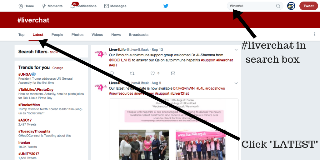
Join HepBUnited, NASTAD, National Viral Hepatitis Roundtable (NVHR) and CDC’s Division of Viral Hepatitis for a Twitter Chat on Hepatitis Testing Day, May 19th at 2 P.M. EDT.  The chat will highlight hepatitis events and allow partner organizations to share their successes, challenges and lessons learned from their efforts, particularly during this unique time. Partners will also highlight innovative strategies for outreach during COVID-19. This twitter chat serves to keep us all informed, raise awareness and share messaging. All are encouraged to join the twitter chat conversation with the hashtag #HepChat20, and to keep partners posted throughout the month about events and messaging with the hashtag #HepAware2020.

Continue reading "Join Hepatitis Partners for a Twitter Chat on May 19th, #HepTestingDay!"

Hepatitis B Transmission for Those Newly Diagnosed

Being diagnosed with hepatitis B can be a confusing experience and may leave you with many questions. Understanding your diagnosis is essential for your health, and understanding how hepatitis B is transmitted can help prevent transmission to others. 

How is it Spread? 

Hepatitis B is transmitted through direct contact with infected blood. This can happen through direct blood-to-blood contact, unprotected sex, unsterile needles, and unsterile medical or dental equipment. Globally, hepatitis B is most commonly spread from an infected mother to her baby due to the blood exchange during childbirth. It can also be transmitted inadvertently by the sharing of personal items such as razors, toothbrushes, nail clippers, body jewelry, and other personal items that have small amounts of blood on them.

Hepatitis B is not transmitted casually by sneezing or coughing, shaking hands, hugging or sharing or preparing a meal.  In fact, hepatitis B is not contracted during most of life’s daily activities. You don’t need to separate cups, utensils, or dishes. You can eat a meal with or prepared by someone with hep B. Hugging, or even kissing won’t cause infection unless there are bleeding gums or open sores during the exchange. As an infection that is spread through the blood, standard precautions such as covering all wounds tightly, practicing safe sex (using a condom), and cleaning up all blood spills with gloves and a solution of one part bleach to nine parts water will protect against transmission. The best tool we have to prevent transmission is the hepatitis B vaccine!

Most of those who are newly infected have no notable symptoms. This is why it is important to encourage family members and sexual partners to get tested if you test positive. Often, it remains undetected until it is caught in routine blood work, blood donation, or later in life after there is liver inflammation or disease progression. 

Dealing with a Possible Exposure:

One important factor for those that may have been exposed is the timing. There is a 4-6 week window period between an exposure to hepatitis B and when the virus shows up in the blood (positive HBsAg test result).  If you go for immediate testing, please understand that you will need to be re-tested 9 weeks later to confirm whether or not you have been infected. It is essential to practice safe sex and follow general precautions until everyone is sure of their status –both the known and potentially infected.

You may still be in a waiting period trying to determine if you are acutely or chronically infected. It is possible that you have not had symptoms with your hepatitis B. It’s also very likely you are unsure  as to when you were infected. Not knowing the details of your infection can be stressful and confusing, but the most important thing to do now is to educate yourself so that you can take the proper steps to protect your liver and prevent transmission. 

Preventing Future Transmission: 

  1. Always cover open wounds. Keep cuts, bug bites – anything that bleeds or oozes – covered with a bandage. It’s also a good idea to carry a spare bandage.  
  2. Be sure to practice safe sex (use a condom) until you are sure your partner has completed their hepatitis B vaccine series. Be aware that multiple sex partners and non-monogamous relationships can expose you to the potential of more health risks and even the possibility of a co-infection, so it is best to use a condom. Co-infections are when someone has more than one serious chronic condition (like HBV and HCV , HBV and HIV or HBV and HDV).  Co-infections are complicated health conditions that you want to avoid. Therefore, practice safe sex by using a latex or polyurethane condom if you have multiple partners.
  3. Keep personal items personal.  Everyday items that are sharp may contain small amounts of blood. This includes things like razors, nail clippers, files, toothbrushes and other personal items where microscopic droplets of blood are possible. This is good practice for everyone in the house. Simple changes in daily habits keep everyone safe!

If a person has been tested and their results show that they are not already vaccinated or have not recovered from a past infection, then they should start the series as soon as possible. This includes sexual partners and close household contacts and family members. The HBV vaccine is a safe and effective 2 or 3-shot series.  

If you wish to confirm protection, the timing of the antibody titre test should be 4-8 weeks following the last shot of the series. If titers are equal to or above 10 mIU/mL, then there is protection for life.  If someone has been previously vaccinated a titer test may show that their titers have waned and dipped below the desired reading. There is no reason to panic, as a booster shot can be administered and then a repeated titer test 1-2 months later can ensure adequate immunity. Once you know you have generated adequate titers, there is no need for concern of transmission!

When recovering from an acute infection, if your follow up blood test results read: HBsAg negative, HBcAb positive and HBsAb positive then you have resolved your HBV infection and are no longer infectious to others and you are no longer at risk for infection by the HBV virus again.

However if your follow up blood tests show that you are chronically infected or your infection status is not clear, you will want to take the precautionary steps to prevent transmitting your HBV infection to others. You will also need to talk to your doctor to be sure you have the appropriate blood work to determine your HBV status and whether or not you are chronically infected.

Please be sure to talk to your doctor if you are unsure, and don’t forget to get copies of all of your lab results!


I had a Liver Transplant Because of Hep B: Here’s What You Should Know

April is Donate Life Month in the United States. Donate Life Month is primarily known as a time to acknowledge those who have saved the lives of others by donating an organ, but it is also a chance to highlight the incredible journeys of those who have required organ transplants. 

Two years ago, Peter V. had a seven-hour emergency liver transplant after a chronic hepatitis B infection led to rapid liver failure. Peter sat down with us and shared an in-depth look into why he needed a liver transplant and how it changed his life. 

  1. Why did you need a liver transplant? 

I had acute liver failure.  About 1 and ½ years before my liver failure, I was taken off the hepatitis B medication (Viread) by my gastroenterologist and to maintain blood work monitoring about every 6 months.  From the span of June of 2017 to January of 2018 immediately before my liver failed, my hepatitis B DNA went from 1,000 IU/L to 169 million IU/L and my ALT went from 24 IU/L to 4,419 IU/L.  By this time, my liver had completely been destroyed through cirrhosis. Without the hepatitis B medication, the virus can flare up at any time and reaction to it once this happens could be too late as in my case.

2. What did a liver transplant mean to you previously? Did you realize how serious the procedure was? 

I never thought about a liver transplant, or any transplant for that matter. I never thought I would need one. Before, life with hepatitis B was normal and routine. Hepatitis B was simply part of my life; I took my medications and had no side effects from them, so the liver transplant was a surprise to me. 

I didn’t understand how serious a transplant was. My condition deteriorated rapidly when my liver began to fail. I couldn’t even do basic functions like unlocking my phone. It got to the point where my situation was so severe that I was in and out of consciousness; I didn’t even know that they were taking me into surgery. Upon recovery, when my cognitive function came back, my wife informed me that my situation had been extremely critical. 

3. What kind of treatment and follow-up did the transplant involve? 

I was bed-ridden and unconscious for over two weeks after the transplant. I needed physical therapy to regain my strength –  to sit up or to get out of bed. For two weeks, I had therapy three or four times a day to regain my ability to speak and cognitive thinking. 

Blood work is also a big part of follow-up. In the beginning, I had to have my blood taken daily before it lessened to once a week, then once every two weeks, and eventually to once a month. All of the blood tests are to make sure that your body does not reject the new organ. I’m on anti-rejection medications, but there is always the risk that your body can reject it. About a year ago, my ALT number rose to high levels, which raised immediate concern. My post-transplant team took a sample of my liver and found that my body was rejecting the new liver. They increased my anti-rejection medications and my body was able to adapt. 

I also developed diabetes after my transplant and had to be placed on insulin, however, I was able to stop taking it by changing my diet and monitoring my blood sugar through my eating habits. 

4. How has the liver transplant changed your life? Are you still able to carry out daily activities the same way you did previously?

I don’t drink alcohol at all anymore and I take much better care of my body. Before my transplant, I didn’t take hepatitis B seriously. I was still drinking alcohol and wasn’t eating a healthy diet. The transplant made me realize how serious hepatitis B could be if you don’t take care of your liver. 

The anti-rejection pills suppress my immune system, which means I have to be very careful about what I eat and how it is prepared. Eating out at restaurants is a risk because you have to trust that the restaurants are properly cleaning their food and that it is cooked properly.  Because of the immunosuppressants, improperly cleaned food can be dangerous. One time I developed a fever and had to be hospitalized because of cross-contamination between foods at a restaurant. You really have to be aware of what you are eating. 

The reminders for COVID-19 to wash your hands thoroughly and not touch your face have been my life since the transplant. Eventually, these actions become a habit. I am still able to work and do physical activities like yard work and exercise, but it is not as vigorous as before. I still get fatigued throughout the day, but it doesn’t stop me from living my day-to-day life. 

I don’t know how I would do this without support. I don’t know how I would do this by myself. I still do physical therapy to regain my strength and my family helps while I go through this journey. Family support is key. I also have a post-transplant team that will help monitor my health for at least 3 years after my transplant.

Most importantly, my family support has been abundant and going through an experience like this makes me much more appreciative of the love and care from having family support that should not be taken for granted. Now, the time spent together regardless of the activities are much more precious.

5. Is there anything that you wish you could have changed about your experience? 

I wish I didn’t have to go through the transplant. I wish I knew more about how serious hepatitis B was. I still drank alcohol and ate the same foods that I ate before my diagnosis. I neglected my liver health. I wasn’t serious about it before the transplant; hepatitis B was invisible to me. It shouldn’t have taken a liver transplant for me to become aware of it. 

6. What have you learned since your journey? Do you have any advice for those living with hepatitis B who think that a liver transplant is the best, or only, option for them?

I don’t think that a liver transplant is an “option”. For me, it was life-or-death. Because I was in critical condition, I was able to get it immediately. For others, getting a transplant is a long and difficult journey. 

A liver transplant is not going to get rid of chronic hepatitis B. You will still live with it. Hepatitis B is still a part of my life every day. The difference is that I now have an understanding of what it can do to my liver. 

Having chronic hepatitis B is not life-ending. It’s not even life-changing as long as you take your medication and take care of your liver. I put my friends and family through a scary experience. If you have chronic hepatitis B, take your medications and keep your liver healthy. Take your diagnosis seriously. 

About Liver Transplants for Those Living with Hepatitis B:

A liver transplant is a very serious surgery that removes a diseased or injured liver and replaces it with a healthy one. People living with hepatitis B can need a liver transplant if their liver begins to fail. This typically occurs if the infection is not being monitored properly, or if significant liver damage has been occurring. Regular monitoring by a knowledgeable provider, a healthy lifestyle, and taking medications, if needed, as prescribed, can help prevent the need for a liver transplant.

 Thank you, Peter, for providing a look into your experience! 

Hepatitis B Research Review: February


Welcome to the Hepatitis B Research Review! This monthly blog shares recent scientific findings with members of Baruch S. Blumberg Institute (BSBI) labs and the hepatitis B (HBV) community. Technical articles concerning HBV, Hepatocellular Carcinoma, and STING protein will be highlighted as well as scientific breakthroughs in cancer, immunology, and virology. For each article, a brief synopsis reporting key points is provided as the BSBI does not enjoy the luxury of a library subscription. The hope is to disseminate relevant articles across our labs and the hep B community. 

 Summary: This month, researchers in Beijing, China have reported that a therapeutic vaccine composed of polylactic acid microparticles loaded with HBV surface antigen and the mouse STING agonist DMXAA showed efficacy in clearing HBV infection in a mouse model. Researchers from Wuhan, China have reported that SOX2, a transcription factor important for cell proliferation is also a host restriction factor for HBV infection. Also, researchers from the University of Boulder in conjunction with Dr. James Chen’s lab in Dallas have reported the synthesis of two potent cGAS inhibitors.

The incorporation of cationic property and immunopotentiator in poly (lactic acid) microparticles promoted the immune response against chronic hepatitis B – Journal of Controlled Release

This paper from the Chinese Academy of Sciences in Beijing, introduces a microparticle vaccine which may be used to treat chronic HBV infection (CHB). The 1μm diameter microparticle is made from polylactic acid (PLA), which is a biodegradable polymer typically synthesized from plant starch. The microparticle also contains didodecyldimethylammonium bromide (DDAB) which is a double-chain cationic surfactant. This group has previously shown that DDAB may be used as a carrier for the HBV surface protein (HBsAg). DDAB also gives the microparticle a positive charge, which accelerates its phagocytosis into antigen-presenting cells (APCs) and facilitates its escape from lysosomal degradation once in the cell. Additionally, the group loaded microparticles with the mouse STING agonist  5,6-dimethylxanthenone-4-acetic acid (DMXAA). The microparticles were refereed to as DDAB-PLA (DP) and DDAB-PLA-DMXAA (DP-D) respectively. Both types of microparticle were saturated with HBV surface antigen (HBsAg). The microparticles were first tested on mouse bone marrow dendritic cells (BMDCs). Administration of microparticles caused less than a 20% reduction of cell viability in these cultures. BMDCs treated with DP-D microparticles had at least ten-fold more expression of IRF-7 and IFN-β mRNA as measured by RT-qPCR than those treated with HBsAg or DP microparticles alone. Surprisingly, the DP-D microparticle-treated cells also had about twice the expression of these genes compared to the positive control HBsAg + DMXAA, which contained ten times more DMXAA than the microparticles. This indicates that the DP-D microparticles induced the STING pathway with high efficiency due to their bioavailability. Next, the group found that DP-D microparticles induced the highest level of chemokine expression (measured via RT-qPCR) and immune cell recruitment (measured via flow cytometry) at the site of injection in inoculated mice compared with HBsAg alone, HBsAg with aluminum salts (traditional vaccine adjuvant), and DP microparticles. This result shows that the DP-D microparticles induced both an innate immune response and an adaptive immune response in mice. Further, the group showed that BMDCs treated with DP-D microparticles had a high level of maturation, expressing CD40, CD86, and MHCII molecules on their surface as measured by flow cytometry. Finally, the group administered the HBsAg-primed microparticles to mice infected with recombinant HBV (rAAV-1.3HBV virus, serotype ayw). Mice treated with both types of microparticles showed a higher cytokine response as well as a higher titer of anti-HBsAg antibody as measured by ELISA. Mice treated with the DP-D microparticles had the most profound immune cell activation and  fastest clearance of serum HBsAg. The microparticle vaccine introduced in this publication is promising because it is highly efficient in delivering antigen to immune cells. The microparticles are unique in that they contain a small molecule STING agonist inside. This design is clever because this vaccine stimulates the innate immune system by activating STING and the adaptive immune system by displaying HBsAg to APCs. This promotes HBV clearance in a multifaceted approach: immune cells produce cytokines through the STING pathway, T cells recognize and destroy infected cells, and B cells secrete anti-HBsAg antibodies to neutralize newly formed viruses. This publication highlights the versatility of biodegradable microparticle technology in designing unique approaches to combat infection. Micro- and nanoparticle delivery systems represent a promising avenue for future drugs to combat HBV and other viruses.

SOX2 Represses Hepatitis B Virus Replication by Binding to the Viral EnhII/Cp and Inhibiting the Promoter Activation – Viruses
This paper from Wuhan University in China identifies the protein sex determining region Y box 2 (SOX2) as a host factor that restricts HBV replication. SOX2 is a transcription factor critical for cell proliferation and the tumorigenecity of solid tumors. In 2006, expression of SOX2 along with three other transcription factors was shown to convert somatic cells into induced pluripotent stem cells. Overexpression of SOX2 indicates poor prognosis in patients undergoing resection of HCC. In HCC cells, SOX2 has also been found to induce the expression of programmed death ligand-1 (PD-L1), leading to the tumor’s evasion of the host immune system. Previously, it has been demonstrated that HBV infection induces increased expression of SOX2 in hepatocytes. This study demonstrates that SOX2 inhibits HBV replication by binding to the Enhancer II (EnhII) and Core Promoter (Cp) regions of the HBV genome. By binding to the EnhII/Cp region, SOX2 disrupts the transcription of the mRNA species precore, core, polymerase, and pgRNA. This reduction of mRNA transcription results in reduced levels of core-associated DNA, HBV surface antigen (HBsAg), and HBV e antigen (HBeAg). To learn this, the group co-transfected both HepG2 and Huh7 cells with a fixed concentration of  HBV 1.3-mer plasmid DNA alongside variable concentrations of Flag-tagged SOX2 in pcDNA3.1 plasmid DNA. Cells transfected with higher concentrations of SOX2 plasmid DNA showed reduced levels of HBV mRNAs (3.5, 2.4, and 2.1 kb) via Northern blotting. SOX2-transfected cells also showed reduced levels of HBV core-associated DNA via qPCR as well as reduced levels of both HBsAg and HBeAg via ELISA. Next, in order to learn  if SOX2 interacts directly with an HBV promoter, a dual-luciferase reporter assay was implemented. Here, four vectors were used, each containing one of the HBV enhancer and/or promoter sequences (preS1, preS2, EnhⅡ/Cp, and EnhⅠ/Xp) upstream of a firefly luciferase reporter. Each of these firefly luciferase reporter vectors were co-transfected into HepG2 cells alongside variable concentrations of SOX2 plasmid DNA. A plasmid encoding Renilla luciferase was also included at a constant concentration in each transfection as a control for transfection efficiency. While firefly luciferase has an emission of 625 nm (red), Renilla luciferase has an emission of 525 nm (green). Therefore, levels of red fluorescence were used to measure the activity of the HBV enhancer/promoter sequences and levels of green fluorescence were utilized as a control for transfection efficiency. Co-transfection with SOX2 significantly diminished the luciferase activity of the EnhII/Cp reporter only and in a dose-responsive manner, indicating its interaction with that region of the HBV genome. Further, using HBV-producing HepAD38 cells, chromatin immunoprecipitation coupled with quantitative PCR (ChIP-qPCR) was used to isolate SOX2 protein and then determine what DNA sequence it was bound to. The EnhII/Cp sequence was found to be highly enriched on SOX2 protein. In order to determine which part of the SOX2 protein is required for binding to the EnhII/Cp region, truncated forms of SOX2 were generated in the pcDNA3.1 plasmid. Using the assays described above, it was found that only SOX2 mutants lacking the high mobility group (HMG) domain were unable to bind to the EnhII/Cp region and suppress HBV products. Interestingly, it was found that SOX2 mutants lacking the transcription activation (TA) domain were still able to bind to the EnhII/Cp region. Further, it was demonstrated by Western blot of subcellular fractions and immunofluorescence that SOX2 mutants lacking the HMG domain were unable to enter the nucleus. Finally, studies were performed in an in vivo BALB/c mouse model. Mice were given a hydrodynamic injection of an adeno-associated viral vector conferring HBV (pAAV-HBV1.3) alongside pcDNA3.1 plasmid DNA conferring SOX, SOX2 lacking HMG domain ( SOXΔHMG), or empty vector. Levels of HBsAG and HBeAg in the blood at days two and four were reduced only in mice given the full length SOX2 plasmid. Additionally, mice given the full length SOX2 plasmid had a reduction of 3.5kb HBV mRNA in liver tissues as measured by qPCR and a lower abundance of HBV core antigen (HBcAg) in liver tissues as measured by immunohistochemical staining. This study shows that SOX2 protein, previously shown to be upregulated by HBV, plays an anti-HBV role in the liver. SOX2 is therefore a new host restriction factor of HBV replication. SOX2 may be one protein which contributes to HBV-induced hepatocarcinogenesis, given its role in promoting the transcription of genes involved in cell proliferation. In the future, SOX2 may be utilized for its anti-HBV activity or targeted for the treatment of HCC.

 Discovery of Small Molecule Cyclic GMP-AMP Synthase Inhibitors – The Journal of Organic Chemistry

This paper from the University of Colorado Boulder introduces the development of novel small molecule inhibitors of the protein cyclic GMP-AMP synthase (cGAS). This publication is in conjunction with Dr. James Chen’s laboratory at the University of Texas Southwestern Medical Center in Dallas, Texas. Dr. Chen’s lab discovered cGAS in 2012. cGAS is a cytosolic, double-stranded DNA (dsDNA)-sensing protein. It belongs to the nucleotidyltransferase family of enzymes which transfer nucleoside monophosphates, the substituents of nucleic acids. When cGAS recognizes dsDNA, it synthesizes the cyclic dinucleotide cyclic GMP-AMP (cGAMP). cGAMP acts as a second messenger and activates the stimulator of interferon genes protein (STING). Once activated, STING triggers TBK1- and IKK-mediated activation of the transcription factors interferon regulatory factor 3 (IRF3) and nuclear factor kappa-light-chain-enhancer of activated B cells (NF-kB). In the nucleus, IRF3 and NF-kB induce the expression of type I interferons and other inflammatory cytokines. cGAS is essential for detecting foreign pathogens which contain dsDNA and triggering an innate immune response to clear them. However, excessive or dysfunctional cGAS activity may lead to chronic inflammation and/or autoimmunity. Pharmacologic inhibition of cGAS may provide treatments for diseases including Aicardi-Goutiés syndrome (AGS), lupus erythematosus, and cancer. Current small molecule inhibitors of cGAS are limited by poor specificity and/or cellular activity. In this study, a high throughput virtual screen (HTVS) was utilized to screen about 1.75 million drug-like compounds for activity against the dimer-forming and DNA-binding faces of mouse cGAS (mcGAS). mcGAS was utilized for the in silico screen because the human cGAS (hcGAS)-DNA complex was only recently published. From this virtual screen, ten compounds were further investigated, leading to the selection of one lead compound. This lead was further optimized for greater potency through chemical modifications resulting in the analogues CU-32 and CU-76. The IC50 of both compounds is below 1µM. To test these compounds’ selectivity for cGAS, human monocyte cells THP-1 were either transfected with  interferon-stimulatory DNA (ISD) or infected with Sendai virus (SeV). ISD is a 45-basepair DNA known to activate cGAS, while SeV is a single-stranded RNA (ssRNA) virus known to activate the RIG-I-MAVS pathway; both stimuli are known to result in IRF3 activation and dimerization. Following treatment with both compounds, Western blot of the cells was conducted probing for the formation of IRF3 dimers. In ISD-treated cells, CU-32 and CU-76 inhibited the formation of IRF3 dimers in a dose responsive manner. Neither compound had any effect on IRF3 dimer formation in SeV-infected cells. This result indicates that these inhibitors are selective to cGAS. Using in silico molecular docking studies, the group speculates that these compounds disrupt the interface of the cGAS dimer, allosterically inhibiting dimerization. The discovery of novel cGAS inhibitors is exciting and important for multiple reasons. These compounds, if made commercially available will allow for improved experimentation investigating the cGAS/STING pathway. If these compounds or their derivatives are found to be safe and effective in humans, they may be promising candidates for the treatment of autoimmune disorders or cancer.


Meet our guest blogger, David Schad, B.Sc., Junior Research Fellow at the Baruch S. Blumberg Institute studying programmed cell death such as apoptosis and necroptosis in the context of hepatitis B infection under the direction of PI Dr. Roshan Thapa. David also mentors high school students from local area schools as part of an after-school program in the new teaching lab at the PA Biotech Center. His passion is learning, teaching and collaborating with others to conduct research to better understand nature.


Hepatitis B Research Review – February

Welcome to the Hepatitis B Research Review! This monthly blog shares recent scientific findings with members of Baruch S. Blumberg Institute (BSBI) labs and the hepatitis B (HBV) community. Technical articles concerning HBV, Hepatocellular Carcinoma, and STING protein will be highlighted as well as scientific breakthroughs in cancer, immunology, and virology. For each article, a brief synopsis reporting key points is provided as the BSBI does not enjoy the luxury of a library subscription. The hope is to disseminate relevant articles across our labs and the hep B community. 

This paper from the University of Duisburg-Essen in Germany shows that hepatocytes infected with HBV exhibit innate immune signaling via the pattern precognition receptor (PRR) Toll-Like Receptor 2 (TLR2). The adaptive immune response to HBV infection is well characterized and is broken into phases based on serological testing of antibodies produced against the virus. However, whether HBV infection triggers an innate immune response has remained controversial, with the long-held belief being that HBV evades the innate immune system as a “stealth virus”. Contrary to this view, studies of acute HBV infection in patients have indicated an early, innate immune response to HBV characterized by a natural killer (NK) cell response. Toll-like receptors (TLRs) are a class of membrane-bound receptor proteins which play a key role in innate immunity by recognizing foreign pathogens and activating inflammatory signaling cascades. A previous publication from this group has demonstrated that primary human hepatocytes (PHHs) can be stimulated through the TLR proteins TLR1-9. In this paper, PHHs from human donors were infected with HBV ex vivo. Then, expression of the innate immune cytokines Interleukin 1 Beta (IL1B), Interleukin 6 (IL6), and Tumor Necrosis Factor Alpha (TNFα) were measured by quantitative, reverse-transcription polymerase chain reaction (qRT-PCR). HBV-infected PHHs showed greatly increased expression of these genes at three hours after infection compared to mock-infected and not treated PHHs. Additionally, immunocytochemical staining revealed translocation of the transcription factor nuclear factor kappa-light-chain-enhancer of activated B cells (NFκB) to the nuclei of HBV-infected PHHs, indicating a cytokine response. Next, to characterize the innate immune response caused by HBV infection, a DNA microarray was used. Here, PHHs were either infected with HBV or treated with a known TLR ligand such as Pam3Cys (TLR2 agonist) or poly(I:C) (TLR3 agonist). Then, RNA was extracted from the cells and converted through a complementary DNA (cDNA) intermediate into biotin-labeled anti-sense RNA (aRNA) which was then hybridized to a Human Genome U219 Array Plate. This plate, coated with over 530,000 DNA probes representing over 20,000 human genes served as a scaffold for complementary base-pair binding of the aRNAs derived from the cells. Once bound to the microarray, the biotin-labeled aRNAs were detected by staining with streptavidin phycoerythrin, resulting in a fluorescent signal wherever complementary base-paring occurred. This microarray analysis revealed which specific inflammatory genes were up-regulated in the PHHs by each stimuli. Gene expression signals which were induced by HBV infection were compared with those induced by the TLR agonists. The gene expression profile of HBV-infected PHHs was most similar to that of PHHs treated with the TLR2 agonist Pam3Cys. This data indicates that HBV infection induces a TLR2-like innate immune response. Importantly, no expression of interferon-stimulated genes (ISGs) was detected in the microarray analysis. Finally, PHHs were pre-treated with neutralizing antibodies against TLR2 (nABTLR2) prior to infection with HBV. HBV-mediated induction of IL1B, IL6 and TNF was significantly reduced by nABTLR2 pre-treatment and conversely, HBV replication was increased. In summary, this paper shows that PHHs exhibited an innate immune response to HBV infection via the TLR2 pathway. The group suggests that this response is one of the body’s first steps leading to HBV clearance. Furthermore, in the discussion section the group indicates that the HBV surface antigen (HBsAg) is likely the protein component of HBV which activates TLR2 upon infection. This finding may help in the development of strategies to cure chronic HBV infection.

​This paper from Wuhan University in China reports that HBV infection can increase the expression of Programmed Death Ligand 1 (PD-L1) on the surface of infected hepatocytes, allowing them to escape destruction by the adaptive immune system. PD-L1 is the binding partner of Programmed Death 1 (PD-1), an immune checkpoint protein on the surface of T cells. The expression of PD-L1 on cell surfaces allows for their recognition by circulating T cells as part of the body and not an outside threat. This interaction is important for the prevention of autoimmune disorders in which the immune system attacks healthy cells of the body. However, PD-L1 is commonly over-expressed in a number of cancers and is a hallmark of especially aggressive cancers. PD-L1 expression on cancer cells allows them to neutralize T cells which specifically target them. This is one example of an “immune-escape” strategy exhibited by cancers. Accordingly, PD-L1 and PD-1 are the target of a number of FDA approved immunotherapies for cancer including the PD-L1 inhibitors Tecentriq, Bavencio, and Imfinzi and the PD-1 inhibitors Keytruda, Opdivo, and Libtayo. These drugs are some of the first in their class in that they are not small molecules, but are recombinant, monoclonal antibodies. Phosphatase and tensin homologue deleted on chromosome 10 (PTEN) is a tumor suppressor which is mutated or deleted in many human cancers. PTEN is a phosphatase, a protein which dephosphorylates other molecules. This group has previously shown that PTEN plays a role in antiviral innate immunity. Therefore, they wanted to see if PTEN also regulates the adaptive immune response in the context of HBV infection. First, they used immunohistochemical staining of patient liver tissues to compare the levels of PTEN and PDL-1 in patients chronically infected with HBV vs healthy controls. There was a reduced staining of PTEN and a heightened staining of PD-L1 in chronic HBV tissues compared to controls. The group then found a similar correlation using immunofluorescence, qPCR and Western blotting of HepG2 cells vs HepG2.2.15 (HBV-producing) cells. They also transfected HepG2 cells and infected mice via hydrodynamic injection with an HBV-containing vector (pHBV1.3) or an empty vector control (pUC18) and then performed qPCR and/or Western blotting.  In all systems, HBV infection/production induced a reduction of PTEN and an increase in PD-L1 expression. Then, in order to elucidate this phenomenon further, a PTEN-expressing plasmid was transfected into HepG2.2.15 cells, which resulted in a reduction in PD-L1 mRNA and protein. Conversely, PTEN knockdown in HepG2.2.15 cells resulted in a two-fold increase in PD-L1 mRNA and protein expression. These results show that HBV inhibits PTEN expression which in turn causes up-regulation of PD-L1. Next, the group transfected HepG2 and Huh7 cells with a number of constructs conferring individual HBV proteins. They found that HBV X protein (HBx) and HBV polymerase (HBp) reduced PTEN expression more than any other HBV protein components. Next, the group analyzed how HBV production in hepatocytes affected human T cells grown in co-culture. Jurkat T cells were co-cultured with either HepG2 or HepG2.2.15 cells and then analyzed by flow cytometry. Jurkat T cells grown alongside the HBV-producing HepG2.2.15 cells had a higher incidence of apoptosis, a higher expression of PD-1, and less Interleukin-2 (IL-2) secretion than those grown alongside HepG2 cells. This result indicates that HBV-infected hepatocytes suppress local T cell responses by PD-L1/PD-1 signaling. Finally, the group used a mouse model of HBV infection to show that PTEN over-expression promotes HBV clearance in vivo. This paper shows that PD-L1, a highly studied drug target implicated in the immune-escape of cancers is also up-regulated by HBV infection. Furthermore, the HBV proteins responsible for this up-regulation are HBx and HBp. This finding may help in the development of  immunotherapies to treat chronic HBV infection. Perhaps FDA approved PD-L1 or PD-1 inhibitors may be used in conjunction with interferon alpha treatment or HBV antivirals to boost the immune response against HBV-infected hepatocytes.

This paper from National Tsing Hua University in Hsinchu, Taiwan reports the design and testing of nanoparticles which selectively confer immunogene therapy to hepatocellular carcenoma (HCC) cells. Nanoparticles are very small (1-1000nm) particles which have become an attractive novel drug candidate in recent years. The use of nanoparticles as medicine would enable the customizable delivery of DNA, RNA, or protein payloads to cells. The novel nanoparticles presented here deliver both a small interfering RNA (siRNA) against the Programmed Death Ligand 1 (PD-L1) gene as well as a plasmid DNA (pDNA) encoding the cytokine Interleukin 2 (IL-2). The strategy behind the nanoparticles’ design is to both inhibit an immunosuppressive gene (PD-L1) and up-regulate an immunostimmulatory gene (IL-2) in tumor cells. Delivery of such genes to tumor cells would make them more vulnerable to destruction by circulating cytotoxic T cells (CD8+ T cells). This type of approach is needed, because many advanced tumors create an immunosuppressive tumor micro-environment (TME) rendering many cancer treatments ineffective. The nanoparticles presented here are referred to as tumor-targeted lipid dendrimer-calcium phosphate (TT-LDCP) nanoparticles. The nanoparticles consist of a core of calcium phosphate, thymine-capped polyamidomine (PAMAM) dendrimers, siRNA, and pDNA. This core is coated with an inner lipid called DOPA and outer leaflet lipids DOPC, DOTAP, and DSPE-PEG. The nanoparticle is then tagged with SP94 (SFSIIHTPILPL), a polypeptide which selectively binds to HCC cells but not healthy hepatocytes. Dendrimers are repeatedly-branching molecules which exhibit a sphere-like shape. PAMAMs are the most well-characterized class of dendrimers, consisting of branching amide and amine groups. The calcium phosphate and PAMAM dendrimers in the core of the TT-LDCP nanoparticle promote endosomal escape of the nucleic acid payload. Additionally, this group shows that the PAMAM dendrimers in TT-LDCP nanoparticles also activate the STING pathway. The group showed that STING was activated by treating mouse HCC cells HCA-1 with complete nanoparticles or those lacking the dendrimers. Cells treated with complete nanoparticles showed, by Western blot a higher level of both TBK1 and IRF3 phosphorylation than those treated with incomplete nanoparticles. Those cells treated with complete nanoparticles also displayed heightened transcription of the STING-triggered proinflammatory genes Ifnb,Ccl5, and Cxcl10 as measured by qPCR. Furthermore, the group showed that treatment using their nanoparticles of mice bearing orthotopic HCC implants resulted in dendritic cell maturation in those animals, regardless of the identity of the genes delivered. These results indicate that the dendrimers used in the TT-LDCP nanoparticles not only serve for efficient delivery of nucleic acids, but also as adjuvants that stimulate the STING pathway and activate tumor-infiltrating dendritic cells. This publication gives a glimpse into what future therapies for cancer may look like. The nanoparticle designed by this group is unique in that it has multiple functionalities: selectively targeting HCC cells, inhibiting PD-L1 expression, inducing IL-2 expression, and activating the STING pathway. Such a complex design is bound to require fine tuning before it can become a medicine. But a multi-target immunotherapeutic such as this may be exactly what is needed to help the body fight against aggressive, immunosupressive tumors.

Lay Summary: 
This month, the innate immune system was the focus of HBV research. Scientists hope to find how the innate immune system interacts with HBV during viral infection and proliferation. Doing so will shed light on host factors which lead to chronic infection and inform antiviral strategies. Notably, this month a human protein, MX2 was found to have potent anti-HBV activity by preventing cccDNA formation. Also, a microRNA encoded by HBV called HBV-miR-3 was found to activate the human innate immune system to limit HBV replication. This month, a paper studying woodchuck hepatitis virus (WHV) traked activation of the innate immune system as well as he adaptive immune system in an acute infection model. Also this month, concerning hepatocellular carcenoma (HCC), the alternative splicing of mRNA in tumors was found to vary in HCC patients based upon their risk factor (HBV, HCV, or alcohol). Finally, a review was published this month concerning STING, an innate immune protein which is not activated by HBV infection but which may prove a valuable tool for cancer treatment.  

Meet our guest blogger, David Schad, B.Sc., Junior Research Fellow at the Baruch S. Blumberg Institute studying programmed cell death such as apoptosis and necroptosis in the context of hepatitis B infection under the direction of PI Dr. Roshan Thapa. David also mentors high school students from local area schools as part of an after-school program in the new teaching lab at the PA Biotech Center. His passion is learning, teaching and collaborating with others to conduct research to better understand nature.